Light manipulation of transient phases in quantum materials is becoming an increasingly popular method for creating novel properties and functions, such as creating nanoscale topological defects. However, visualizing the formation of a new phase in a solid is still difficult due to the vast variety of spatial and temporal scales associated with the process.

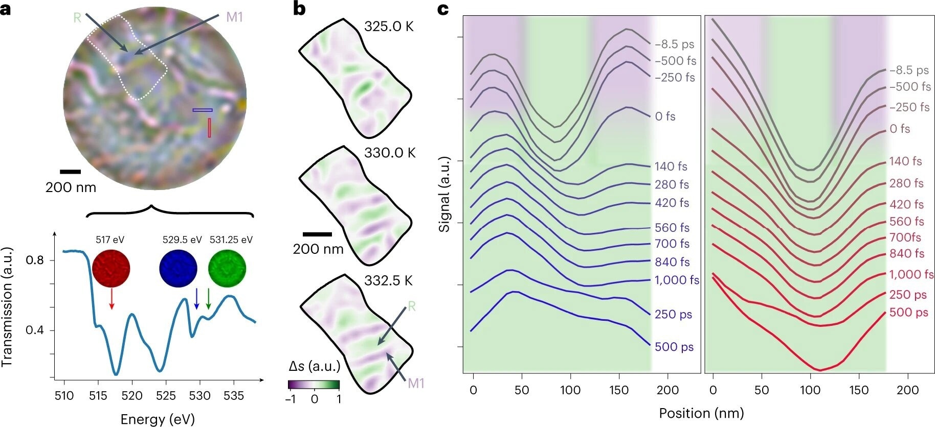
Time-dependent X-ray holographic imaging of VO2. a, False color composite FTH image of VO2 from images recorded on the VO2 soft X-ray resonance (bottom) at 517 eV (red), 529.5 eV (blue) and 531.25 eV (green). The metallic R phase appears green and insulating M1 phase appears purple. b, Temperature-dependent domain growth highlighted through the subtraction of the blue and green channels, Δs, which removes the sample morphology. The region of interest used is indicated by the white dotted region in a. c, Transmission dynamics of two line-outs spanning R regions surrounded by the M1 phase. Their positions are indicated in a and color-coded. The domain structure, initially ~50 nm, is promptly lost. Background is shaded according to state of the material as a guide to the eye. Credit: Nature Physics (2022). DOI: 10.1038/s41567-022-01848-w
Recently published in Nature Physics, researchers from ICFO collaborated with teams from Aarhus University, Sogang University, Vanderbilt University, the Max Born Institute, the Diamond Light Source, ALBA Synchrotron, Utrecht University, and the Pohang Accelerator Laboratory to introduce a new imaging method that enables the light-induced phase transition in vanadium oxide (VO2) to be captured with high spatial and temporal resolution.
Optically Driven Phase Transition in Vanadium Oxide
The phase transition in vanadium oxide (VO2) upon exposure to light has garnered significant attention due to its potential applications in optically driven quantum substances.
At ambient temperature, VO2 exists in the monoclinic insulating (M1) phase, characterized by forming dimerized pairs of vanadium ions. However, upon exposure to light, the M1 phase can undergo a rapid transition to the high-temperature rutile metallic (R) phase.
This transition has been the subject of extensive study and has contributed to developing several innovative techniques, including the use of time-resolved X-ray diffraction and ultrafast X-ray absorption, to understand the nature of the transition.
It has been observed that the transition from the M1 to the R phase involves the disordering of vanadium pairs on a sub-100 femtoseconds (fs) timescale, accompanied by the collapse of the bandgap on a similar timescale.
While much progress has been made in understanding this process, it remains an open question whether the collapse of the bandgap occurs before or as a result of the structural transition to the R phase.
Non-Equilibrium Phases in Transient State Dynamics
Previously, it has been observed that heterogeneity in the transient state plays a significant role in the dynamics of the process under investigation. An analysis of terahertz conductivity indicates that a rutile metallic phase nucleates and grows locally between tens and hundreds of picoseconds.
Moreover, electron diffraction results suggest the existence of a meta-stable heterogeneous monoclinic metallic phase that may persist for hundreds of picoseconds or even microseconds.
This phase differs from the transient phase that appears in the first 100 femtoseconds and is maintained by a structural deformation that maintains monoclinic symmetry. However, the presence of these non-equilibrium phases is still debatable since a direct observation of the metastable monoclinic metallic state is yet to be established.
Highlights of the Current Research
In this study, the researchers used time- and spectrally-fixed resonant soft X-ray coherent imaging to study the light-induced phase transition at the nanoscale with ultrafast temporal resolution.
This approach, which may provide a comparison between distinct phases as well as retrieve quantitative spectrum data for phase recognition, was applied in two modes: Coherent diffractive imaging (CDI) and Fourier transform holography (FTH).
In FTH, scattering signals are immediately inverted using a fast Fourier transform and only need a single observation, allowing for faster data compilation but at the risk of losing the quantitative measurements of the complicated transmission.
CDI, on the other side, employs numerous exposures to expand the dynamic spectrum of observed scattering signals, with images acquired by recurrent phase retrieval methods that reveal the sample's quantifiable transmissions
New X-ray imaging technique to study the transient phases of quantum materials
Video Credit: Science X: Phys.org, Medical Xpress, Tech Xplore/YouTube.com
Important Findings of the Study
The researchers could better grasp the kinetics of the phase change in VO2 due to this novel technique. It was discovered that pressure has a much more significant influence on light-induced phase transformations than previously predicted.
"We saw that the transient phases aren't nearly as exotic as people had believed! Instead of a truly non-equilibrium phase, what we saw was that we had been misled by the fact that the ultrafast transition intrinsically leads to giant internal pressures in the sample millions of times higher than atmospheric. This pressure changes the material properties and takes time to relax, making it seem like there was a transient phase," says Allan Johnson, a postdoctoral researcher at ICFO and lead author of the study.
The results of the study have two main implications. Firstly, there is no evidence to support the existence of a heterogeneous monoclinic metallic phase. The strained orthorhombic state is distinct from previously proposed out-of-equilibrium monoclinic phases, which do not consider strain effects and result from decoupling internal degrees of freedom.
However, strain effects may be responsible for the long-lasting diffraction features that have been associated with these phases. Additionally, the absence of nucleation and growth of the rutile metallic phase indicates that previously observed dynamics could be due to a higher resistivity of strained VO2 than the fully relaxed state.
The findings of this study highlight the crucial importance of obtaining spatially and spectrally resolved measurements to fully comprehend and interpret the transient phases of quantum materials. This novel approach can also be applied to studying filament growth under electric fields, enabling spectroscopy of field-induced states.
Reference
Johnson, A. S. et al. (2022). Ultrafast X-ray imaging of the light-induced phase transition in VO2. Nature Physics. Available at: https://doi.org/10.1038/s41567-022-01848-w
Disclaimer: The views expressed here are those of the author expressed in their private capacity and do not necessarily represent the views of AZoM.com Limited T/A AZoNetwork the owner and operator of this website. This disclaimer forms part of the Terms and conditions of use of this website.