Nanoparticles (NP) are often used in the medical field as a way to deliver drugs to the body. However, when these nanoparticles come into contact with biological environments, they form a protein corona.

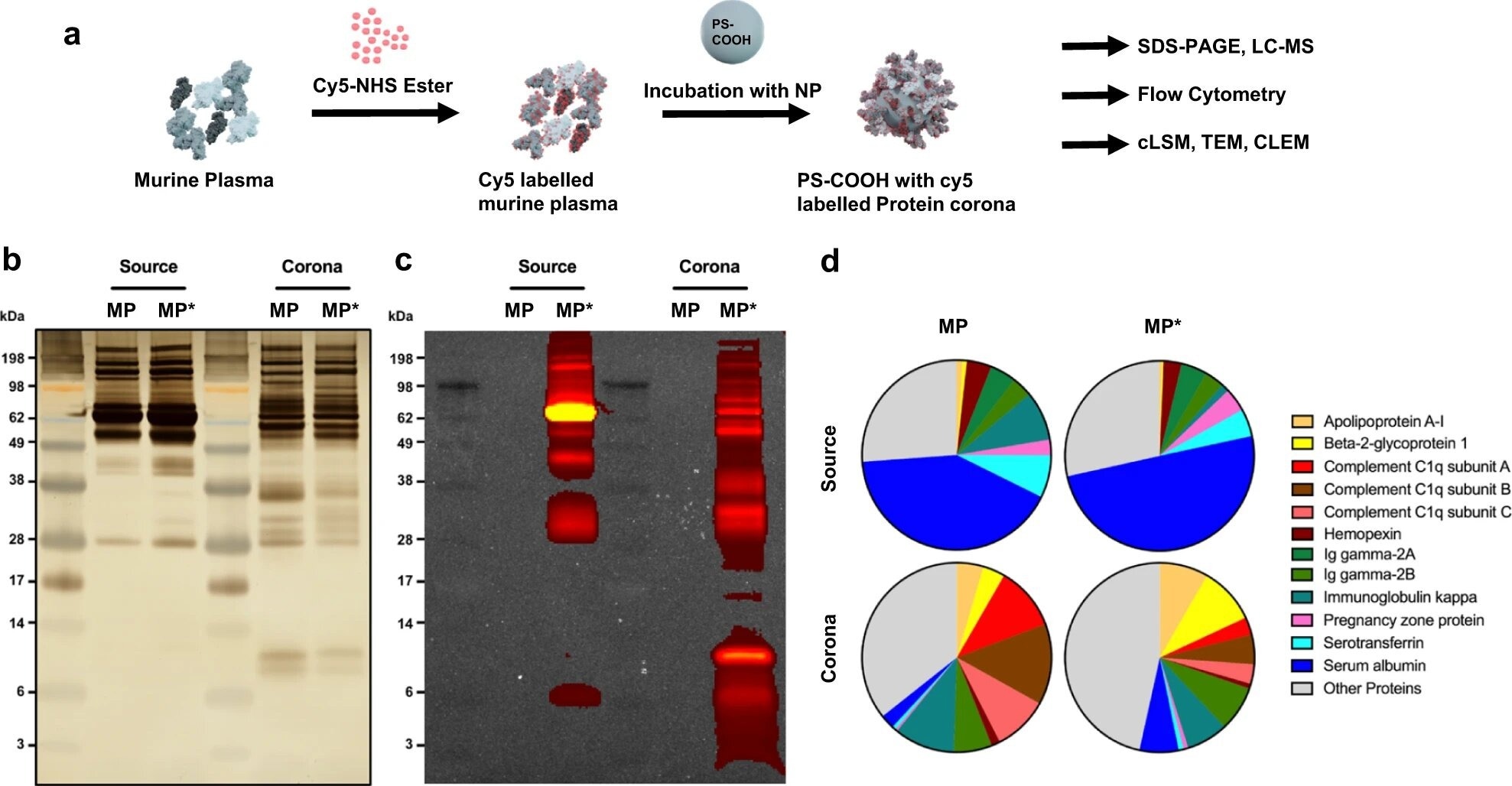
Protein analysis of unlabeled and Cy5-labeled murine plasma and protein corona. a Murine plasma proteins were labeled with Cy5 by NHS-chemistry. Carboxyl-functionalized PS NPs were incubated in unlabeled and Cy5-labeled murine plasma, respectively, to form a protein corona. b Unlabeled murine plasma (MP), Cy5-labeled murine plasma (MP*), and associated protein corona samples were analyzed by SDS-PAGE and silver staining. Corona proteins were obtained after incubation of carboxyl-functionalized PS NPs in plasma, washing, and desorption with 2% of SDS. c Analysis of in-gel fluorescence. The Cy5-fluorescence was imaged by IVIS at an excitation wavelength of 640 nm and an emission wavelength of 680 nm. d Quantitative LC-MS proteomic analysis. The pie charts display the proteins with at least 4% presence in the proteome. Values are represented as the percentage based on all identified proteins. Credit: Nature Communications (2023). DOI: 10.1038/s41467-023-35902-9
While much research focuses on the effects of protein corona creation extracellularly or the repercussions of absorption, very little is understood about the destiny of the protein corona within cells.
A recent study published in the journal Nature Communications aims to bridge this knowledge gap by following the path of such nanoparticles into a cell using a combination of several microscopy methods.
Nanoparticles for Drug Delivery: Overview and Significance
Nanoparticles are a promising tool for delivering drugs to the body because of their unique properties. The small size of NPs allows them to pass through cell membranes and reach specific cells or tissues in the body that would otherwise be inaccessible. This targeted delivery increases the potency of drugs and reduces the side effects.
A protein corona is a coating of biomolecules that forms on the surface of nanoparticles when they come into contact with biological environments. The coating, also called a protein shell, is composed of proteins, lipids, and other biomolecules that adsorb to the surface of the NPs.
The formation of a protein corona is a well-known effect in nanomedicine, as it plays a crucial role in determining the fate of NPs within the body. The composition of the protein corona can change the way NPs interact with cells, which can impact their circulation, uptake, toxicity, and the release of the active pharmaceutical ingredient (API) from the NPs.
Challenges in Understanding the Path of Protein Corona
One of the key challenges in the field of nanomedicine is understanding the fate of the protein corona on the surface of the NPs when they come into contact with biological environments. The protein corona is a coat of biomolecules that adsorbs to the surface of the NPs, altering the chemical identity of the NP and impacting the way it interacts with cells.
While researchers hope to create the protein corona in a controlled manner, exactly what happens to the protein corona after a nanoparticle is taken up by a cell remains unclear. Furthermore, the intracellular destiny of protein corona on nanoparticles is critical for understanding and developing any medication delivery strategy, making this lack of awareness a key research area.
The formation of protein corona is also a dynamic process that can change over time, which poses additional challenges in understanding the behavior of NPs in biological environments.
Highlights of the Current Study
In this study, researchers used correlative light and electron microscopy (CLEM), a fluorescence staining method coupled with electron microscopy. This method is effective for investigating the subcellular destination of NPs and the protein corona.
The technique allows for imaging of the NP and protein corona in the same sample using both light and electron microscopy, thus providing detailed information on the ultrastructure of the NPs and protein corona.
The researchers used a specific fluorescence staining technique to label the NPs and protein corona, which enabled them to track the localization of the NP and protein corona in the cell. The staining technique was combined with electron microscopy to obtain high-resolution images of the NPs and protein corona in different cellular compartments.
This approach allowed the researchers to study the fate of the NP and protein corona at the ultrastructural level, providing new insights into the intracellular fate of protein corona on nanoparticles.
Findings and Future Perspective
In this study, the research team observed the separation between the nanoparticles and the protein corona as they were internalized in different cellular compartments. For instance, the cell directs the nanoparticles towards recycling endosomes, whereas the protein corona gathers in multivesicular bodies.
A separation between the NP and protein corona suggests that the cell has a mechanism for sorting and directing the NPs and protein corona to different cellular compartments. The team concluded that this separation was to prepare the nanoparticles for subsequent exocytosis while the protein corona remains in the cell to be metabolized.
"We, therefore, assume that the drug release in the cell is not disturbed by the protein coating," says Ingo Lieberwirth, co-author of the study. "However, it is now important to find out how exactly the process takes place inside the cell."
The study reported the intracellular fate of the protein corona on NPs for the first time, providing new insights into the behavior of nanoparticles in biological environments. In the future, this knowledge could be used to develop strategies for controlling the formation and properties of the protein corona to achieve tailored effects.
Reference
Han, S. et al. (2023). Endosomal sorting results in a selective separation of the protein corona from nanoparticles. Nature Communications. Available at: https://doi.org/10.1038/s41467-023-35902-9
Disclaimer: The views expressed here are those of the author expressed in their private capacity and do not necessarily represent the views of AZoM.com Limited T/A AZoNetwork the owner and operator of this website. This disclaimer forms part of the Terms and conditions of use of this website.