Jul 18 2008
What is there to see inside a magnet that's smaller than the head of a pin?
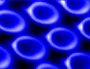
 Image of an array of microscopic magnets taken with scanned probe ferromagnetic resonance force microscopy -- a new imaging technique invented by Ohio State University physicists and colleagues. The disk-shaped magnets measure only two micrometers (millionths of a meter) across. Image courtesy of Ohio State University.
Quite a lot, say physicists who've invented a new kind of MRI technique to
do just that.
The technique may eventually enable the development of extremely small computers,
and even give doctors a new tool for studying the plaques in blood vessels that
play a role in diseases such as heart disease.
In a recent issue of Physical Review Letters, the scientists report the first-ever
magnetic resonance image of the inside of an extremely tiny magnet.
Specifically, the magnet is a "ferromagnet" -- a magnet made of ferrous
metal such as iron. It's what most people think of when they hear the word "magnet."
"The magnets we study are basically the same as a refrigerator magnet,
only much smaller," said project leader Chris Hammel, Ohio Eminent Scholar
in Experimental Physics at Ohio
State University. The disk-shaped magnets in this study measured only two
micrometers (millionths of a meter) across.
"Because ferromagnets generate such strong magnetic fields, we can't study
them with typical MRI. A related technique, ferromagnetic resonance, or FMR,
would work, but it's not sensitive enough to study individual magnets that are
this small."
Likewise, medical researchers can't use MRI to image plaques formed in the
body, because plaques are too small. That's why this new kind of magnetic resonance
could eventually become a tool for biomedical research.
The technique combines three different kinds of technology: MRI, FMR, and atomic
force microscopy.
They dubbed the technique "scanned probe ferromagnetic resonance force
microscopy," or scanned probe FMRFM, and it involves detecting a magnetic
signal using a tiny silicon bar with an even tinier magnetic probe on its tip.
As the probe passes over a material, it captures a bowl-shaped image: a curved
cross-section of an object. The magnetic signal is more intense in the middle
(the "bottom" of the bowl), and fades away toward the edges.
It may sound like an odd configuration, but that's why the new technique works.
Every atom emits radio waves at a particular frequency. But to know where those
atoms are, scientists need to be able to localize where the radio waves are
coming from.
Large-scale MRI machines, such as those in hospitals, get around this problem
by varying the magnetic field by precise amounts as it sweeps over an object.
The computer controlling the MRI knows that where the magnetic field equals
X, the location equals Y. Sophisticated software combines the data, and doctors
get a 3D view inside a patient's body.
For Hammel's tiny magnets, no methods were previously known that would image
the inside of them, much less allow for precise localization. But since the
new probe system generates a magnetic field that varies naturally, the physicists
discovered that they could sweep the probe over an array of magnets and get
a 2D view that's similar to a medical MRI. In Physical Review Letters, they
reported an image resolution of 250 nanometers (billionths of a meter).
Now that they have their imaging technique, Hammel and his team are beginning
to record the properties of many different kinds of tiny magnets -- a critical
first step toward developing them for computer memory.
Experts believe that one day, tiny magnets could be implanted on a computer's
central processing unit (CPU) chip. Because system data could be recorded on
the magnets, such a computer would never need to boot up. It would also be very
small; essentially, the entire computer would be contained in the CPU.
For biomedical research, the technique could be used to study tissue samples
taken from plaques that form in brain tissues and arteries in the body. Many
diseases are associated with plaques, including Alzheimer's and atherosclerosis.
Currently, researchers are trying to study the structure of plaques in detail
to understand how they form and how they affect conventional MRI images.
Hammel and his team hope to contribute to the development of an instrument
that could be sold and used routinely in laboratories. But the technique needs
some further development before it could become an everyday tool for the computer
industry or for biomedicine.
Hammel's Ohio State coauthors on the paper include Yuri Obukhov, a research
associate; Thomas Gramila, associate professor of physics; Denis Pelekhov, a
research scientist in the university's Institute for Materials Research; Palash
Banerjee, a postdoctoral researcher; and Jongjoo Kim and Sanghun An, both doctoral
students. They collaborated with Ivar Martin, Evgueni Nazaretski and Roman Movshovich
of Los Alamos National Laboratory; and Sharat Batra of Seagate Research, the
research and development center of hard drive manufacturer Seagate Technologies.
This work was funded by the Department of Energy.