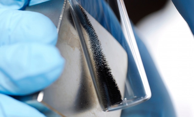
Magnetic nanoparticles are a group of nanoparticles that can be manipulated with a magnetic field. These particles commonly comprise magnetic particles like nickel, cobalt, iron and their chemical compounds. Nanoparticles are normally smaller than 1 µm in diameter and the larger microbeads are 0.5–500 µm in diameter. Magnetic nanoparticles have been researched extensively since they have attractive characteristics that could be used in catalysis, specifically nanomaterial-based catalysts. They have been investigated for use in the following applications:
- Magnetic resonance imaging
- Biomedicine
- Magnetic particle imaging
- Data storage
- Nanofluids
- Environmental remediation
- Optical filters
Medical Applications of Magnetic Nanoparticles
Magnetic nanoparticles are used in a wide range of medical applications that include the following:
- A very special feature of magnetic nanoparticles is that they react to a magnetic force and this is used in applications such as bioseparation and drug targeting including cell sorting.
- These nanoparticles have become highly popular due to their use as heating mediators for cancer therapy and for magnetic resonance imaging (MRI).
A class of cationic magnetic nanoparticles, magnetite cationic liposomes can be used as carriers for the introduction of magnetite nanoparticles into target cells as their positively charged surface and negatively charged surface interact, also they can be used in hyperthermic treatments.
- Antibody-conjugated magnetoliposomes commonly known as AMLs are also used in hyperthermic treatments and enable tumor-specific contrast enhancement in MRI through systemic administration. The feature of manipulating cells labeled with magnetic nanoparticles using magnets finds application in tissue engineering. This is possible as magnetic nanoparticles are attracted to a high flux density.
- MCLs and magnetic force was used to build multilayered cell structures and a heterotypic layered three-dimensional coculture system.
It is anticipated that applying the unique features of these functionalized magnetic particles will enhance medical techniques.
X-rays and MRIs are truly a breakthrough achievement in the field of medicine; however, very soon it will be possible to regulate physiological and molecular changes taking place in the body. Dark brown oxide nanoparticles are anticipated to significantly improve the capabilities of presently available medical imaging techniques.
These nanoparticles, normally less than 50 nm in diameter, comprise an iron oxide core covered by an organic shell. Due to their magnetic nature, these particles are known as superparamagnetic iron oxide (SPIO) nanoparticles.

Magnetic nanoparticles have many promising applications in medicine, including advanced bio-imaging and cancer treatment. Image Credits: Georgia Tech
Enhanced Magnetic Resonance Imaging (MRI)
An MRI scanner includes a huge magnet of 1.5 to 7 Tesla strength and this type of imaging does not use radiation. Radio waves and a magnetic field are used to excite the protons in the body. After excitation, these protons relax and this data is translated into cross-sectional pictures of human tissue by a computer program.
According to an assistant professor at the University of Texas in Dallas, Michael Biwer, a main aspect of present imaging techniques is not just that water is present but also there is a change in relaxivity of water molecules close to the metal. This means that the relaxation rate of the excited water molecules is highly significant for imaging. Diseased and normal tissues offer different relaxation rates that can be read by the MRI image.
Light and dark contrast images are formed on the computer that show a range of relaxation rates. For instance, body areas with higher water content appear brighter in contrast to denser areas. The higher the contrast; the more the resolution of the computer image. Wherever the nanoparticles aggregate in the body, a clear black-and-white contrast is sensed. The strong magnetic characteristics of the SPIO magnetic nanoparticles helps overcome the poor sensitivity of present contrast agents by generating a higher contrast between the background and the target.
For example, consider a patient undergoing a routine breast cancer scan. Magnetic SPIO particles could be injected into her blood stream to confirm the presence of cancer cells in her breast. Since the tumor has leaky blood vessels, the cancer cells are confronted by the SPIO particles and bind to the cancer cells by a "lock and key mechanism". An excellent contrast image will be formed by the MRI scanner that will help visualize the location and presence of the tumor.
Targeted Cancer Treatment
According to researchers from Korea, it is possible to use nanoparticles as a remote-controlled magnetic death switch to destroy cancer cells from Korea. Jinwoo Cheon and Jeon-Soo Shin of Yonsei University, in Seoul, and their colleagues have attached zinc-doped iron oxide magnetic nanoparticles to an antibody, which targets a receptor on colon cancer cells. The cancer cell’s death receptor DR4 is targeted by the antibody when triggered by a magnetic field and programmed cell death or apoptosis is caused.
According to the researchers, nanoparticles enable control of cell signaling pathways. Cell signaling allows exchange of information between cells and underpins growth metabolism, differentiation and several other processes. It also enables cells to trigger apoptosis to make sure tissues do not grow out of control or cells malfunction.
Adding a magnetic element allowed remote control of the signaling process using a magnetic field. This enables controlling the position of magnetic nanoparticles in the body and specific malignant tissues are targeted. The research team demonstrated this technique using zebra fish and showed that the death switch can be operated at the micrometer scale. Shin emphasized that this is the first time an in vivo magnetic nanoswitch has been demonstrated and been proved feasible.
Conclusion
Magnetic nanoparticles find extensive use in medical applications. It is possible to make chemical modifications to these particles so that they are injectable, non-toxic, compatible with the body and can be concentrated at a high level in the target organ or tissue. Since these particles are magnetic they can be used as contrast agents for MR imaging. With advanced development of these agents, even very small amounts will be enough to render significant contrast between good and tumor tissue.
Researchers are striving to design magnetic nanoparticles that do not just locate diseases and tumors but also treat them by providing precise drug doses. The days are not far off when we will walk into a clinic, the doctor will inject SPIO nanoparticles, the disease will be detected, treated with nanoparticles and we can walk out of the room in a few hours.
Sources and Further Reading