In this interview, AZoNano talks to Dr. Sonja Stözle-Feix, director of scientific affairs at Nanion, and Dr. Mattias Goßmann, CEO of InnoVitro, about the abilities and applications of Nanion’s FLEXcyte technology.
Could you please give us an overview of Nanion Technologies’ main products to date?
Dr. Sonja Stözle-Feix: For around 18 years our core technology has been automated patch clamping. We started with the Port-a-Patch, and now have the Port-a-Patch Mini, which is available since last year. Here, the amplifier is integrated into the system and thus the system offers fast and easy access to high-quality patch-clamp data with only minimal effort.
We also have higher throughput systems, Patchliner and SyncroPatch 384i, with the latter offering up to 768 recordings in parallel. Our portfolio also includes bilayer recording technology, the Orbit family, with which you can measure single-channel events of reconstituted proteins in the lipid bilayers. The SURFE2R technology can do recordings of electrogenic transporters, and then, of course, we have the CardioExcyte and the FLEXcyte technology.
Could you talk us through the capabilities of the CardioExcyte system?
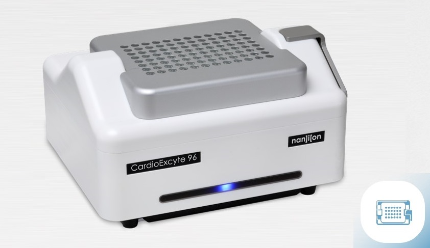
Dr. Sonja Stözle-Feix: The CardioExcyte system comes with a controlled environmental chamber and sits outside of the incubator. It utilizes 96-well consumables, that are long-term stable, and the electrode design allows you to do impedance and EFP recordings with the same cells.
The system also comes with an optical and electrical stimulation capability. The impedance mode allows you to not only look at contractility of cardiomyocytes, for example but also to look at proliferating cells, like cancer cells or hepatocytes. The system comes with a liquid handling robot to help out with cell seeding, compound application, or medium exchange.
Putting all this together allows the system to be a perfect tool for cardiac safety screening. In terms of hardware and software capabilities, the CardioExcyte’s basic system allows you to do impedance and EFP measurements of the same cells in one well. it In some cases, it’s a true advantage to look at both of these signals from the same cell.
If you look at both signals, you do capture the real and true compound effect: For example, compounds de-coupling contraction from the electrophysiology, like Myosin II blockers, can be effectively investigated.
The graphical user interface allows you to capture with one glance your 96 wells and the beating shape or the impedance traces of your experiment. A mean beat is calculated by averaging an individual recording sweep. The advantages are basically that you have a higher signal-to-noise ratio and the calculation of parameters is more consistent.
By selecting the mean beat view, you get the information from the entire well, and you don't have to pick a golden electrode, furthermore, you do have a better overview of your experiment.
For example, if you have cellrhythmia happening introduced by the application of dofetilide, you get an arrhythmic mean beat. You can easily capture this by looking at the graphical user interface and you can judge at a glance if the compound is affecting your cells or not. There are quite a lot of parameters captured while the experiment is going on, during our so-called “online analysis”.
Base impedance and beat rate or beat rate regularity are recorded, and we also capture the primary and secondary beats, which show you arrhythmic effects, pulse width, amplitude, and so on. Similar parameters are also captured in these EFP traces, along with the beat rate, beat rate regularity, amplitude, FPDzero and FPDmax values, and more.
How does CardioExcyte’s software improve data analysis?
Dr. Sonja Stözle-Feix: When analyzing the data, we have a nice piece of software called DataControl. In principle, with DataControl, you can pick any parameter that you like and calculate either IC 50, or EC 50, or percentage of block. In general, DataControl is a software that we make available for different platforms that we have in our portfolio. CardioExcyte and SyncroPatch 384i are also the SURFE2R 96SE system. Furthermore, the license provided with our platform is not user limited. Analysis templates can be stored and shared to ensure consistency, and DataControl96 can be installed on as many computers as you need, which makes it easy to share data and allow certain users to analyze or look at the data.
How have physiological cell responses and historical contractility measurement techniques led to the creation of FLEXcyte technology?
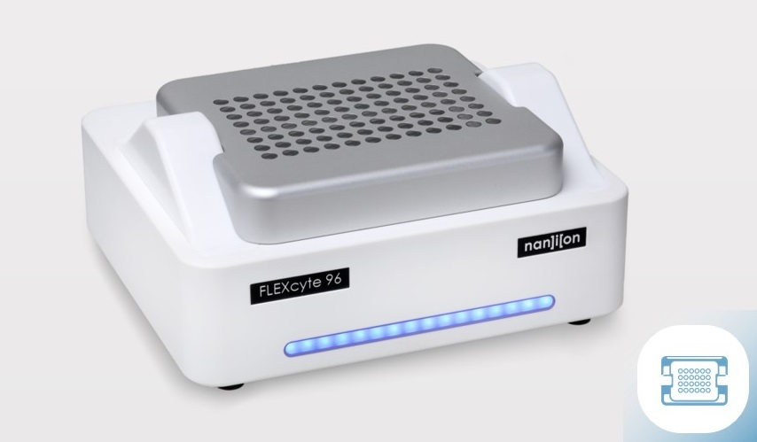
Dr. Sonja Stözle-Feix: The FLEXcyte is an add-on for the CardioExcyte system. The two recording modes can be easily exchanged by the user, simply by exchanging the lid of the device.

FLEXcyte 96
Mattias Goßmann: The state-of-the-art technique when it comes to contractility measurements is still the Langendorff heart and related ex vivo techniques. The Langendorff heart was first described in 1895, but it's still the gold standard. This is despite the fact that it has a list of severe drawbacks, like low predictivity from using animal models over human models, and that it has a very low throughput and is very labor-intensive as you can only use one organ per experiment.
Even so, this technique does have a big advantage, and that is its physiologic response. We believe that this derives from the natural mechanical environment that the cells have. If we look at the tissue level, different things can happen to the cardiomyocytes depending on the actual state of their environment.
If you imagine that you have your cell in a very soft substrate like a gel, the cardiomyocyte can contract one time and it will contract maximally. That is what we call isotonic contraction. The other extreme is an isometric contraction, where the cell has infinite rigidity of its surrounding environment. That's exactly what happens if you seed the cells on a very hard substrate, like glass or plastic. This is where we have this maximum stress that we go through with each cycle of contraction or relaxation, and the cardiomyocytes are going from one extreme to the other.
But, under physiological conditions, we have auxotonic contractions. The cells have this certain kind of mechanical resistance in their surroundings and their environment, and this withstands the contraction and makes a positive feedback loop between force, stress, tension, and mechanical resistance. The cells above that can actively sense what's happening in their surroundings.
Flexcyte 96 - True contractility
In short, cardiomyocytes do sense their surroundings by the use of deformable adaptor proteins. The protein that they use the most is actin, and they use it to sense fast muscle myosin contraction and slow non-muscle myosin contraction.
The deformation of different adaptor proteins regulates the differential protein kinase pathways; if the surrounding is too stiff, the actin will be stretched too much, and if it's too soft, the actin won’t be stretched enough. Above that, they are mechanosensitive on a gene expression level. Cardiomyocytes respond to their physiologic matrix stiffness with drastic transcriptional deregulation.
With cells on soft and medium substrates and polystyrene, a large portion of genes is deregulated. But on very soft gels, it’s been found that only seven genes were deregulated, and it was mainly metabolic processes that were affected. Often we consider the cytoskeleton or other processes to be relevant, but it’s also metabolic processes that determine the fate of a cell in a significant way.
Ion channels of cardiomyocytes also influence mechanosensitivity, and some studies have shown that cells are stimulated by stretching and their calcium influx increased with increasing magnitude of stretching. It was found that if RNA was silenced in L-type calcium channels, which are some of the most important channels in cardiomyocytes, a large portion of those channels won’t work properly on very rigid substrates if they are stretch sensitive.
The basic challenges that we have if we come to the market and want to replace animal-based ex vivo models with human stem cell-derived in vitro models are, of course, the productivity and the physiological relevance. But additionally, what we see regularly is that we have to increase our throughput quite a lot. We also need to be very cost-effective.
These principles are what led to this technology, and we call it FLEXcyte technology. So this is an add-on to the CardioExcyte, but in this case, the cells are cultured on ultra-thin silicon substrates with adjustable mechanical environments. Those substrates with the cells on top of it are deflected downwards by the weight of the medium on top of it so that whenever the cells contract simultaneously, the membrane is lifted upwards by a degree. When they relax, it goes down again. Their cellular stress can be calculated from the deflection.

CardioExcyte 96
What are the main differences between the impedance-based systems and the FLEXcyte system?
Mattias Goßmann: The main parameters that are different are due to the substrate. On impedance-based systems, you have an infinitely stiff substrate, so the contraction that the cells can exert there are isometric contractions that are not physiological. The parameter is the dynamic change in cellular shape.
With the FLEXcyte, we have very physiologic stiffness and, consequently, we have an auxotonic contraction. That's also the basic parameter that we measure: the contraction properties of the cells.
The electronics are very similar, just that we use impedance to measure real contraction. The amplitude is a very important parameter for contractility, but it's not the only parameter that is of importance.
We also have the durations and downstrokes. We have the corresponding slopes of those phases, and of course, the integrals. We can also measure frequency and arrhythmic events.
What are the main advantages of the FLEXcyte system?
Mattias Goßmann: We believe that the FLEXcyte system provides additional physiological relevance as cardiomyocytes can sense the mechanical environment that they would sense in the human body, and their structures and their parts can work as they should. We have quite a high throughput with 96 wells and quite a good cost-effectiveness.
Additionally, the FLEXcyte directly integrates into Nanion's accessories, especially the temperature control and CO2 incubation system. You do not have to operate your system in the incubator, but you can operate it as a benchtop device. It also integrates into an SOL head for optical placing. This works nicely with the channelrhodopsin-2, which is useful. The FLEXcyte integrates very well into the existing system.
Does the FLEXcyte work with contracting cell types other than cardiomyocytes, e.g. smooth and skeletal muscles?
Dr. Sonja Stözle-Feix: In principle, anything that contracts with a sufficient force could be recorded in the FLEXcyte plates. We are currently working on testing different muscle type cells apart from the heart. Stay tuned for any upcoming applications.
Have you observed any changes in expression patterns of cardiomyocytes when grown on FLEXcyte plates in comparison to stiff substrates?
Dr. Sonja Stözle-Feix: At the moment we are talking mainly about phenotypical responses that we capture, but we hope to get more information on the expression levels and on the protein levels that are expressed in the membranes. It’s an ongoing project.
How does the FLEXcyte compare to 3D models?
Dr. Sonja Stözle-Feix: In principle, by seeding cardiac fibroblasts we can achieve three-dimensional structures. The 2D culture has the advantage that comparably lower cell numbers are required, but when using fibroblasts together with cardiac cells, you could come to a 3D model cell type.
How can our readers learn more about FLEXcyte?
Mattias Goßmann: If readers are interested in using such a device in their lab, they should let us or Nanion know! We’re also planning several demos in the upcoming days and weeks.
About Dr. Sonja Stözle-Feix
 Dr. Sonja Stoelzle-Feix currently serves as the director of scientific affairs at Nanion Technologies, and since 2016 has been the co-chair of the CiPA high throughput screening ion channel working group.
About Dr. Mattias Goßmann
 Dr. Mattias Goßmann is the co-founder and CEO of InnoVitro, and is an expert in biotechnology, and holds a doctorate in chemistry.
Disclaimer: The views expressed here are those of the interviewee and do not necessarily represent the views of AZoM.com Limited (T/A) AZoNetwork, the owner and operator of this website. This disclaimer forms part of the Terms and Conditions of use of this website.