Electron microscopy methods have been workhorse techniques in nanotechnology because their spatial resolution is on the order of nanometers.

Image Credit: PolakPhoto/Shutterstock.com
Few standard optical microscopy methods can achieve the same level of spatial resolution, which is essential for being able to resolve geometric and structural features on the nanometer length scale.
Electron beam microscopy methods work by either passing a beam of electrons through a sample onto a detector, known as transmission electron microscopy, or through the detection of secondary or scattered electrons in scanning electron microscopy. Detection of either kind of electron allows for reconstruction of the size of the imaged object as well as visualization of any more complex structural features.
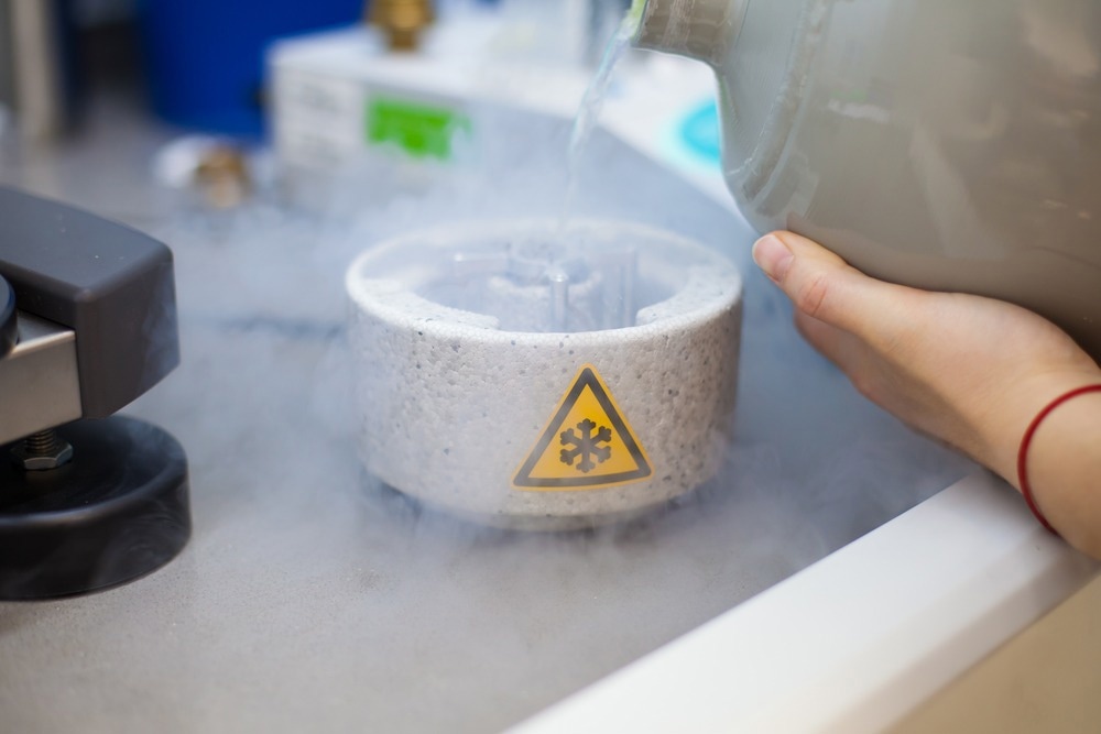
Cryo-electron microscopy (cryo-EM) uses many of the same principles as other electron microscopy methods but differs in sample preparation. In cryo-EM the sample is ‘flash frozen’ using cryogenic liquids such as liquid nitrogen. The flash freezing process provides a high-quality crystalline sample that can then be imaged to recover the typical structural and elemental composition information achievable using electron microscopy.
The advantage of cryo-EM approaches over conventional electron microscopy is that it is possible to retrieve the types of structural information that would normally be obtained using X-ray crystallography methods for which it is very difficult or impossible to grow high-enough quality crystals to perform crystallography experiments.
The flash freezing ensures the size of any crystals formed is minimal and does not disrupt the overall structure. Once the sample has been frozen, it can be added to the electron microscopy grid for measurement.
Another key advantage of cryo-EM is the reduced sample damage. Many of the original developments in cryo-EM were motivated by biological imaging applications. Biological samples can be fragile and easily damaged by the intense electron beams required to produce the brightest images. Finding ways to further reduce the electron beam intensity required for measurements and therefore increase the types of samples that can be studied is a highly active area of research.
Cryo-EM in Nanoscience
Examples of cryo-EM applications in nanoscience include nanoparticle visualization and sizing, imaging of weakly bonded materials that would degrade under standard electron microscopy conditions, and structural nanotechnology.
Improvements in detector sensitivities, in particular direct electron detector sensitivities, meant it is now possible to perform particle sizing with cryo-EM at the single particle level. Improving detection efficiency has also been advantageous for using lower-intensity electron beams for measurement and reducing sample damage.
For biological applications, one of the biggest challenges in cryo-EM has been achieving images with sufficient signal-to-noise as exposure times and beam intensities need to be reduced to avoid sample damage and the resulting image distortion.
Now, with these improvements, it is possible to measure even complex biological nanotechnology systems, including protein-based nanoreactors. By taking small nanoscale structures, such as protein cages, it is possible to use the structure to perform given biochemical or chemical reactions, such as ketene or alcohol reduction and oxidation.
Such reactions can be used to exploit the natural biochemistry of some species, like enzymes and proteins, which tend to be both highly efficient and selective in producing given chemical or biological products.
Many new nanoparticle designs are based on biological structures, such as self-assembled DNA origami or lipids. Polymeric nanoparticles that often contain organic monomeric units have also benefitted from the imaging conditions that can be achieved in cryo-EM.
Improvements in imaging reconstruction methods have also meant full three-dimensional structures can be recovered even for objects with highly complex structures.
Commercial Options
Price has been a prohibitive factor in the adoption of electron microscopy and, in particular, cryo-EM. A standard electron microscopy set-up can already represent a significant investment, with the most basic models starting at many tens of thousands of pounds and more advanced machines costing hundreds of thousands. This price also does not consider any additional costs associated with any data processing or reconstruction architecture that is also required.
As reconstructing three-dimensional structures requires relatively intense computational processing as well as handling large amounts of data, finding more cost-effective and affordable ways to improve this step, including the exploitation of cloud computing resources, is also a highly active area of development.
Several commercial options for electron microscopy are available from companies such as JEOL and Hiatchi. Standard electron microscopy is now a mature technology with many different platforms and imaging options commercially available, including portable or benchtop electron microscopes.
For cryo-EM, the rapid adoption of the technology by the life sciences and nanoscience communities has pushed for the rapid development of commercial options. ThermoFisher offers a number of instrument options for cryo-EM and, where the cost of the cryo-EM platform is prohibitive, or demand is not sufficient, there are a number of companies offering cryo-EM services where samples can be sent for analysis.
Other companies also specialize in selling vitrification kits for the flash freezing of samples, which is an essential part of performing good cryo-EM measurements.
References and Further Reading
Smith, D. J. (2008) Ultimate resolution in the electron microscope? Materials Today, 11, pp. 30–38. https://doi.org/10.1016/S1369-7021(09)70005-7
Callaway, E. (2020) The protein-imaging technique taking over structural biology. Nature, 578, p. 201. https://media.nature.com/original/magazine-assets/d41586-020-00341-9/d41586-020-00341-9.pdf
Dubochet, J., Frank, J., & Henderson, R. (2018) Cryo-EM in drug discovery: achievements, limitations and prospects. Nature Publishing Group, 17(7), pp. 471–492. https://doi.org/10.1038/nrd.2018.77
Baker, L. A., & Rubinstein, J. L. (2010) Radiation damage in electron cryomicroscopy. Methods in Enzymology, 481(C) pp. 371–388. https://doi.org/10.1016/S0076-6879(10)81015-8
De Ruiter, M. V., Klem, R., Luque, D., Cornelissen, J. J. L. M., & Castón, J. R. (2019) Structural nanotechnology: Three-dimensional cryo-EM and its use in the development of nanoplatforms for: In vitro catalysis. Nanoscale, 11(10), pp. 4130–4146. https://doi.org/10.1039/c8nr09204d
Li, Y., Huang, W., Li, Y., Chiu, W., & Cui, Y. (2020) Opportunities for Cryogenic Electron Microscopy in Materials Science and Nanoscience. ACS Nano, 14(8), pp. 9263–9276. https://doi.org/10.1021/acsnano.0c05020
Stewart, P. L. (2016). Cryo‐electron microscopy and cryo‐electron tomography of nanoparticles.pdf. WIREs Nanomed Nanobiotechnol, 9, p. e1417. https://doi.org/doi: 10.1002/wnan.1417
Cianfrocco, M. A., & Leschziner, A. E. (2015) Low cost, high performance processing of single particle cryo-electron microscopy data in the cloud. ELife, 4(MAY), pp. 1–10. https://doi.org/10.7554/eLife.06664
Disclaimer: The views expressed here are those of the author expressed in their private capacity and do not necessarily represent the views of AZoM.com Limited T/A AZoNetwork the owner and operator of this website. This disclaimer forms part of the Terms and conditions of use of this website.