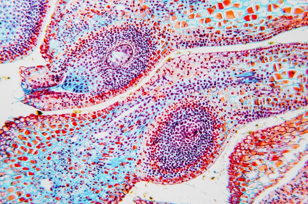
Two-photon microscopy is a type of fluorescence microscopy that, rather than exciting the sample with a single photon, makes use of multiple photons. The advantage over more traditional one-photon methods is that it can be used for applications such as deep tissue imaging.1

Image Credit: Digital Photo/Shutterstock.com
Two-photon microscopy makes imaging at tissue depths of several hundred microns possible, where many standard one-photon methods cannot be used due to very strong light scattering by biological tissues.
A two-photon microscopy measurement uses much of the same instrumentation and procedures as the single-photon equivalent. A light source, typically a laser, illuminates the sample, and fluorescent photons emitted from the sample after photoexcitation are imaged onto a detector. Normally, the sample is on some kind of raster stage, so the full sample can be imaged.
Multiphoton Absorption
The key difference between two-photon microscopy and single-photon equivalent is the intensities of the light used. Focusing a laser beam can be one way to achieve improved spatial resolutions, but for two-photon microscopy, focusing also serves another purpose.
In the case of low light intensities, most samples will only absorb a single photon at a time. There is a linear relationship between the intensity of the light used and the amount of absorption. When the light fields used start to get more intense, particularly if tightly focused femtosecond pulses are used, there is a certain probability of multiple photons being absorbed simultaneously. The intensity dependence of the probability of multiphoton absorption is non-linear. As a result, the feasibility of two-photon microscopy measurements has been heavily dependent on improvements in high repetition rate ultrafast laser systems.2
Sometimes tw- photon microscopy and multiphoton microscopy are used interchangeably as, with sufficiently high peak powers, absorption of three or more photons is also possible.
The absorption of multiple photons results in many differences in the resulting fluorescence image. A continual problem in fluorescence microscopy is the amount of unwanted scattered background light. Confocal methods try to reduce this with the introduction of a confocal pinhole to reject the out-of-focus background but limit the imaging depth. In two-photon microscopy, as only the most intense parts of the laser beam result in two-photon excitation, there is a very small focal volume and, therefore, a high degree of rejection of out-of-focus objects.3
Workflow
The first step of any biological imaging experiment is sample preparation. Sample sections do not need to be as thin as for single photon experiments, but the choice of fluorophore may be different as it is desirable to have fluorophores with large two-photon cross-sections rather than just considering the one photon cross-section.4
Once the sample has been treated and labeled, it can be loaded into the microscope and the two-photon microscopy images can be recorded by rastering the sample under the laser beam. Depending on the instrumentation and level of automation, the user may need to consider checking the focus is optimized and consistent throughout the experiment.
During the experiment, it is necessary to consider photobleaching of fluorophores and whether any sample damage is being caused by the high laser intensities required to drive the multiphoton process. Some optimization of excitation conditions may be required to record meaningful images.
Once the images have been obtained for analysis, interpretation can begin. There have been extensive efforts to develop automated image analysis and recognition algorithms that can automatically identify particular cell structures or species to improve the throughput and accuracy of two-photon microscopy measurements.5
Commercial Market
There are a number of commercial solutions available for two-photon microscopy. Hamamatsu is one example that provides scanning two-photon microscopy measurements. Olympus and Brukker also sell multiphoton microscopy hardware platforms with a number of customizable options depending on the size of samples to be measured and the degree of automation required.
As well as the main microscopy hardware, the two-photon microscopy market also recovers the need for auxiliary reagents for biolabeling and staining as well as specialist laser light sources to generate the intense excitation conditions required.
ThermoFischer Scientific has a number of labels and bioconjugates available for probing different cellular processes in two-photon microscopy, as well as recommendations for filter sets to obtain the best quality images.
Titanium sapphire lasers have been a popular choice for multiphoton microscopy, with their 800 nm fundamental light being a sufficiently long wavelength to avoid excess tissue damage. Coherent and SpectraPhysics both have a range of laser systems optimized for two-photon microscopy applications. Ytterbium-based laser systems, which generally offer better reliability than Ti:sapphire, have become another popular solution, with Light Conversion offering an extensive range of high repetition rate options as well as other devices to allow for wavelength tunability.
Software is another area of active development in the two-photon microscopy market, with a number of solutions becoming available for automated image reconstruction and analysis.
Why is Cryo-Electron Microscopy Used?
References and Further Reading
Helmchen, F., & Denk, W. (2005). Deep tissue two-photon microscopy. Nature Methods, 2(12), pp.932–940. doi.org/10.1038/nmeth818
Phan, T. G., & Bullen, A. (2010). Practical intravital two-photon microscopy for immunological research : faster , brighter , deeper. Immunology and Cell Biology, 88, pp.438–444. doi.org/10.1038/icb.2009.116
Rubart, M. (2004). Two-Photon Microscopy of Cells and Tissue. Circulation Research, 95(12), pp.1154–1166. doi.org/10.1161/01.RES.0000150593.30324.42
Shaya, J., et al. (2022). Design , photophysical properties , and applications of fluorene-based fluorophores in two-photon fluorescence bioimaging : A review. Journal of Photochemistry & Photobiology, C: Photochemistry Reviews, 52(April), p.100529. doi.org/10.1016/j.jphotochemrev.2022.100529
Botez, D., et al. (2018). Quantum cascade lasers. Optical Materials Express, 8(5), pp.1378–1398. doi.org/10.1364/OME.8.001378
Beggs, S., et al. (2015). Applications of the Excimer Laser : A Review. Dermatologic Surgery, pp.1201–1211. doi.org/10.1097/DSS.0000000000000485
Traub, T., et al.. (2014, May). 2.6 um to 12 um tunable ZGP parametric master oscillator power amplifier. In Nonlinear Optics and Its Applications VIII; and Quantum Optics III (Vol. 9136, pp.170-175). SPIE. doi.org/10.1117/12.2052288
Conti, C., et al. (2016). Portable Sequentially Shifted Excitation Raman spectroscopy as an innovative tool for in situ chemical interrogation of painted surfaces. Analyst, 141, pp.4599–4607. doi.org/10.1039/c6an00753h
Li, M., et al. (2022). Integrated Pockels laser. Nature Communications, 13, p.5344. doi.org/10.1038/s41467-022-33101-6
Squier, J., & Muller, M. (2016). High resolution nonlinear microscopy : A review of sources and methods for achieving optimal imaging. Review of Scientific Instruments, 72(7), pp.2855–2867. doi.org/10.1063/1.1379598
Disclaimer: The views expressed here are those of the author expressed in their private capacity and do not necessarily represent the views of AZoM.com Limited T/A AZoNetwork the owner and operator of this website. This disclaimer forms part of the Terms and conditions of use of this website.