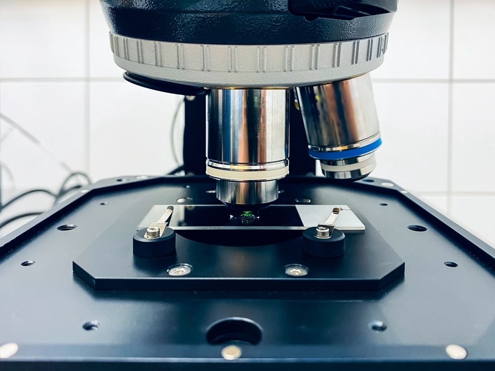
Microscopy techniques have allowed us to see a world that is entirely invisible to our naked eyes. Estimates suggest that the smallest objects resolvable by the naked eye are ~ 0.1 mm – a thousand times larger than even some of the largest nanoscale objects.

Image Credit: Artur Wnorowski/Shutterstock.com
Now, some of the most advanced microscopies are achieving resolutions as high as 7 nm for X-ray methods1 and 0.5 Å for electron microscopies. 2 There has also been a revolution in the resolutions achievable in optical techniques with the development of super-resolution optical microscopy, which can achieve resolutions on the order of sub-100 nm.3 Standard microscopy techniques are limited by Abbe’s diffraction, which has meant previous optical measurements are limited to several hundreds of nanometers.4
In this article, we look at some of the key developments of various microscopy techniques over the last few years and how they have benefitted different areas of research and development.
Electron Microscopy
The impact of an electron microscopy variant, cryo-electron microscopy (cryo-EM), in a field such as electron microscopy was recognized with the 2017 Nobel Prize.5 While electron microscopy methods have been prized for their incredibly high spatial resolutions, one of the difficulties of applying electron microscopy methods to a wider range of materials and biological samples has been the problem of electron damage.
Cryo-EM helped solve the problem of beam damage by improving the damage resistance of samples through cooling. Samples could be flash frozen in a cryogenic liquid, which would freeze them into a particular state, and then be studied with all the advantages of the high structural resolution of electron microscopy methods.
The greater availability of cryo-EM equipment and improvements in measurement protocols and hardware performance have all contributed to making cryo-EM a viable technique for a greater range of samples. Now, cryo-EM is becoming one of the most widely used tools in structural biology, in part because it allows for samples that cannot be crystallized to be measured.6 X-ray crystallography has been one of the main techniques for measuring protein structures but requires good quality crystals to be grown.
Optical Microscopy
Optical microscopy techniques have long been a mainstay in research and medical labs for their versatility and relative simplicity. In recent years, one of the major developments in optical techniques has been the increasing development of automated microscopy platforms that allow for autofocusing and optimization of experimental conditions. Such platforms, combined with advanced image recognition algorithms, are allowing for much higher throughput measurements to accelerate many aspects of drug design and research.7
Super-resolution microscopy methods have seen a huge amount of technical development in the last 20 years.8 Advances in data processing, scanning modalities and a wider range of suitable fluorophores have expanded the range of samples and biological structures that can be measured and the quality of the information that can be obtained.
There are still some challenges in terms of overcoming photobleaching and toxicity issues with some fluorophores and the penetration depth is limited, as is the case with all optical methods, but super-resolution microscopy has still proven invaluable in the analysis of tissue and biological samples.
Some examples of major developments arising from super-resolution microscopy methods include the discovery of how DNA forms nanodomains in its repair mechanisms as well as some of the higher-order structures formed when nucleosomes assemble and how this process occurs.9 Many enzyme structures have now been characterized with super resolution imaging as well as cytoskeletons and subcellular organelles.

Image Credit: Elizaveta Galitckaia/Shutterstock.com
X-Ray Microscopy
One of the key benefits of using X-rays for microscopy, particularly highly energetic X-rays, is that X-rays have significantly greater penetration depths than longer wavelength radiation. The large penetration depth makes X-rays particularly well-suited to imaging either the interior of objects or devices or performing reconstructions of the full three-dimensional object. 3D X-ray microscopy has been used to great effect in the analysis of cellular and sub-cellular details in plant tissues.10
Another interesting aspect of X-ray microscopy methods is the ability to recover information on the elemental composition of the sample. As X-ray radiation interacts with the tightly bound core electrons of an atom, the energetic transitions involved in an X-ray measurement are sufficiently distinctive in energy they are unique to the element being probed.
In X-ray microscopy studies, different images can be reconstructed that visualize where particular elements are localized in a sample, which can be invaluable in understanding how particular species move through a device or object. One example is looking at how particular elements are involved in the degradation processes in batteries and how each has different structural deformations during the degradation process.11
With many synchrotrons moving towards diffraction-limited spot sizes in their upgrade programs, there will be many opportunities for improved spatial resolution in X-ray microscopy.
In conclusion, the variety and accessibility of microscopy techniques have dramatically increased over the last ten years and the growing demand for high-resolution methods in fields such as nanotechnology research will continue to push developments further. Automated image recognition and reconstruction to improve throughput is likely to be another area of microscopy that sees significant investment in research.
References and Further Reading
Rösner, B., et al. (2020). Soft x-ray microscopy with 7 nm resolution. Optica, 7(11), pp. 1602–1608. doi.org/10.1364/OPTICA.399885
Kisielowski, C., et al. (2008). Detection of Single Atoms and Buried Defects in Three Dimensions by Aberration-Corrected Electron Microscope with 0.5-Å Information Limit. Microscopy and Microanalysis, 14(5), pp. 469–477. doi.org/10.1017/S1431927608080902
Huszka, G., & Gijs, M. A. M. (2019). Micro and Nano Engineering Super-resolution optical imaging : A comparison. Micro and Nano Engineering, 2, pp. 7–28. doi.org/10.1016/j.mne.2018.11.005
Nature Photonics Editorial (2009). Beyond the diffraction limit. Nature Photonics, 3, p. 361. doi.org/10.1038/nphoton.2009.100
Shen, P. S. (2018). The 2017 Nobel Prize in Chemistry: cryo-EM comes of age. Analytical and Bioanalytical Chemistry, 410(8), pp. 2053–2057. doi.org/10.1007/s00216-018-0899-8
Dubochet, J., et al. (2018). Cryo-EM in drug discovery: achievements, limitations and prospects. Nature Publishing Group, 17(7), pp. 471–492. doi.org/10.1038/nrd.2018.77
Melanthota, S. K., et al. (2022). Deep learning-based image processing in optical microscopy. Biophysical Reviews, 14(2), pp. 463–481. doi.org/10.1007/s12551-022-00949-3
Bond, C., et al. (2022). Technological advances in super-resolution microscopy to study cellular processes. Molecular Cell, 82(2), pp. 315–332. doi.org/10.1016/j.molcel.2021.12.022
Prakash, K., Diederich, B., & Schermelleh, L. (2022). Super-resolution microscopy : a brief history and new avenues,: Philosophical Transitions A, 380, pp.20210110. doi.org/10.1098/rsta.2021.0110
Duncan, K. E., Czymmek, K. J., Topp, C. N., Jiang, N., & Thies, A. C. (2022). X-ray microscopy enables multiscale high-resolution 3D imaging of plant cells , tissues , and organs. Plant Physiology, 188, pp.831–845. https://doi.org/10.1093/plphys/kiab405
Tsai, E. H. R., Billaud, J., Dario, F., Tsai, E. H. R., Billaud, J., Sanchez, D. F., Ihli, J., Odstr, M., & Holler, M. (2019). Correlated X-Ray 3D Ptychography and Diffraction Microscopy Visualize Links between Morphology and Crystal Structure of Lithium-Rich Cathode Materials Correlated X-Ray 3D Ptychography and Diffraction Microscopy Visualize Links between Morphology and Cryst. IScience, 11, pp.356–365. doi.org/10.1016/j.isci.2018.12.028
Disclaimer: The views expressed here are those of the author expressed in their private capacity and do not necessarily represent the views of AZoM.com Limited T/A AZoNetwork the owner and operator of this website. This disclaimer forms part of the Terms and conditions of use of this website.