Explore how advancements in particle size analysis, including innovative methods like smartphone photogrammetry and artificial intelligence, are transforming industries ranging from pharmaceuticals to environmental protection—continue reading to delve deeper into the topic.

Image Credit: Cergios/Shutterstock.com
A particle is defined as a discrete sub-part of a material. The size of a particle is its most important physical property that directly influences the material properties such as solubility, reactivity, morphology, appearance, viscosity, etc.1 The particle size distribution of a material greatly affects the quality of parts produced by the additive manufacturing process, which has applications in various industries ranging from medicine to aerospace.2
Importance of Particle Size Analysis
Particles are contained in several types of materials. They are found in granules and powders like pharmaceutical ingredients, cement, and pigments. Particles are present in slurries, emulsions, and suspensions like mining mud, milk, and vaccines. Aerosols and sprays, such as inhalers and crop sprays also contain particles of different materials.1 Thus, particles being present in almost everything around us, the particle size analysis becomes a critical parameter for overall material characterization.
In an increasingly competitive and interconnected global economy, product quality control is essential to reap high economic benefits. High-quality products comply well with the safety and performance standards of a regulated market. They allow the manufacturer to charge a higher premium from customers and face low rejection rates.1
A thorough particle size analysis is critical to understanding how it affects the properties of the ingredients and final product. Such particle size clarity allows for the process's optimization to improve product performance. It helps troubleshoot manufacturing and supply issues.1 Apart from individual product improvement, it enhances the robustness of the overall supply chain. Thus, proper particle size analysis can ensure quality control and highly efficient product manufacturing.
Methods for Particle Size Analysis
For simplification, most particle size measurement methods define particle size by the diameter of an equivalent sphere having properties (mass or volume) similar to the original particle. The equivalent sphere models used vary from one technique to another, which explains different results from different analysis methods for the same particle.1
A basic volumetric technique used for particle sizing is sieve analysis.3 During this, the powdered sample is passed through a series of sieves with progressively decreasing mesh sizes. The particle size is concluded to be between the two sieve sizes, the one that the particle fails to pass through and the one just above it through which the particle could pass.2
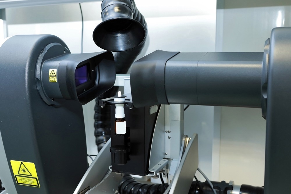
Laser diffraction is widely used for particle sizing in the range of submicrons to millimeters. A laser beam is passed through a dispersed particulate matter, and the angular variations in the intensity of scattered light are measured. Subsequently, the Mie theory of light scattering is used to obtain the particle size distribution. It is a well-established method with high sample throughput due to rapid measurements.1
Dynamic light scattering is a non-invasive technique to measure particle size ranging between submicron to a few nanometers. It applies to particles suspended in a liquid like emulsions, polymers, proteins, and carbohydrates. The suspension is illuminated with a laser, and fluctuations in the intensity of scattered light due to the Brownian motion of particles are recorded. Finally, the Stokes-Einstein equation is used to obtain the hydrodynamic diameter of the particles.1
Automated imaging is another high-resolution technique that directly sizes particles from a few microns to several millimeters. The method involves capturing images of individual particles from the dispersed samples and applies to both stationary samples (static imaging) as well as sample flows (dynamic imaging). It is most suitable for measuring the size of non-spherical particles.1
Scanning electron microscope (SEM) is also commonly used to characterize particles with a size of a few nanometers. It also records 2D images of the particles but provides better resolution than optical sizing methods and is best suited to working on the nanoscale.2
X-ray computed tomography is another image-based method for particle size analysis. However, in contrast to other methods, it captures a full 3D geometry of the particles using X-rays.2
Applications Across Different Sectors
Since particle size influences important properties of a material, the analysis has become an integral part of several key industries; for example, pharmaceuticals (dissolution of tablets, reactivity of catalysts), paints (stability of suspensions), nanomedicines (delivery efficacy), food and beverages (texture and feel), powder coatings and inks (appearance), and ceramics (packing density and porosity).1
Particle size analysis is an important step in various fields of science and engineering. In construction engineering, the particle size distribution of aggregates is a critical factor in determining the strength of asphalt concrete mixtures.
Particle size controls the repose angle of slopes, which is crucial to maintaining the stability of slopes and structures in landslide-prone areas.3 Particle size is also imperative in environmental protection. Effective pollution control cannot be achieved without the size analysis of aerosols and particulate matter.1
Future Trends and Innovations
Even though several techniques are available for particle sizing in a laboratory, efforts are being made to innovate applicable methods on-site. A recent study published in Measurement demonstrated a quick and practical method for particle sizing using smartphone photogrammetry. The researchers created digital elevation models of the particles from the structure captured by motion photogrammetry. The particle sizes thus determined were accurate to a great extent when compared to the sieving method. Since smartphones are available almost everywhere, this rapid, low-cost method can be used for particle size analysis in field conditions.3
Artificial intelligence is the latest technological innovation that is intervening in every field. Particle size analysis is no exception. A recent article in RSC Advances employed deep learning to automate nanoparticle size analysis from secondary electron micrographs (SEMs) and scanning transmission electron micrographs (STEM). The researchers used convolutional neural networks to extract particle size distribution from pseudo-3D images from SEM and STEM. The method has been demonstrated to be highly successful in separating overlapping particles and excluding agglomerates, thus providing accurate particle sizes.4
In conclusion, determining particle size with maximum accuracy and minimum human intervention is the aim of any future developments in this direction. Integration of well-established traditional techniques with new simulation and modeling methods is an expected trend. This will help achieve excellent product performance in the manufacturing industry, effective healthcare for humankind, and efficient environmental protection.
Detecting Defects in Microelectronics Using Particle Analysis
References and Further Reading
- A basic guide to particle characterization. (n.d.). Available at: https://www.cif.iastate.edu/sites/default/files/uploads/Other_Inst/Particle%20Size/Particle%20Characterization%20Guide.pdf
- Whiting, J. G., et al. (2022) A comparison of particle size distribution and morphology data acquired using lab-based and commercially available techniques: Application to stainless steel powder. Powder Technology, 396, pp. 648–662. doi.org/10.1016/j.powtec.2021.10.063
- An, P., et al. (2022) A fast and practical method for determining particle size and shape by using smartphone photogrammetry. Measurement, 193, p. 110943. doi.org/10.1016/j.measurement.2022.110943
- Bals, J., & Epple, M. (2023) Deep learning for automated size and shape analysis of nanoparticles in scanning electron microscopy. RSC Advances, 13(5), pp. 2795–2802. doi.org/10.1039/d2ra07812k
Disclaimer: The views expressed here are those of the author expressed in their private capacity and do not necessarily represent the views of AZoM.com Limited T/A AZoNetwork the owner and operator of this website. This disclaimer forms part of the Terms and conditions of use of this website.