Aug 18 2010
A new lab-based computed tomography (CT) system, capable of delivering synchrotron-like 3D imaging at 50 nanometer resolution within a laboratory setting, was announced today by Xradia, Inc.
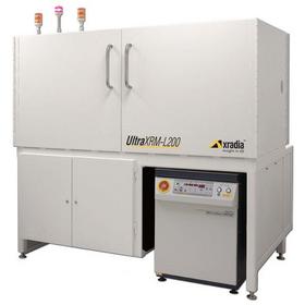
The UltraXRM-L200 is the newest addition to the ultra-high resolution UltraXRM™ nanoscale family of X-ray microscopes. The microscope uses state of the art X-ray optics originally developed for synchrotron research facilities to enable best-in-class resolution and efficiency in lab settings.
 The new UltraXRM-L200 high energy nanoscale X-ray microscope from Xradia: 3D imaging at 50 nanometer resolution for state of the art synchrotron-like capabilities in the lab.
"Our commitment is to continually develop systems that move research forward," said Wenbing Yun, Ph.D., founder, president and CTO of Xradia. "Ultimately, in lab settings as well as at synchrotron facilities, where we have a strong leadership position, we want to help researchers focus on their research rather than invest their time and resources into building their own tools. Only Xradia offers X-ray CT microscopes at this resolution level commercially."
Xradia's UltraXRM-L200 microscope combines a high-flux laboratory X-ray source with proprietary X-ray optics into a standalone CT scanner. It addresses a growing range of applications that include advanced materials development, life science studies for soft tissue and bone, rock porosity studies for oil and gas drilling feasibility models, and semiconductor package failure analysis.
According to Dr. Ge Wang, Professor and Director of the Biomedical Imaging Division at Virginia Tech University, "Xradia's ultra-high resolution imaging systems provide us with detailed 3D volumetric data of the internal structures without the need for cutting or sectioning at the region of interest. This extends the capability of any research laboratory as we are able to test results under a variety of conditions, including time-lapsed 4D imaging. The UltraXRM-L200 will help bridge the gap between existing high resolution imaging modalities such as SEM, TEM and AFM, to optical microscopy and traditional microCT imaging systems."
From Synchrotron to Lab
The only instrument in its class, the UltraXRM-L200 enables timely and efficient synchrotron-like measurement results on demand, without the lengthy delays generally incurred when applying for research time at one of the approximately 50 synchrotron X-ray research facilities in the world.
With a resolution as fine as 50 nm, the UltraXRM-L200 provides insight into microscopic structures and processes previously not accessible with conventional lab-based X-ray technology. Operating with 8 keV (kiloelectron volt) X-rays, it enables observation of structures and materials in their natural state.
Unique capabilities of the UltraXRM-L200
- Non-destructive imaging at such high resolution in a lab system is unique to Xradia, and allows repeated imaging of the same sample under different conditions and/or over time, to bring the fourth dimension -- time -- to analysis;
- Best image contrast: absorption and Zernike phase contrast modes allow imaging of a wide variety of sample types, from rocks to advanced materials to soft tissue;
- Versatility: a large working distance and atmospheric sample environment allow in-situ studies;
- Highest resolution for X-ray CT system: two magnifications, 65 micron field of view at 150 nm resolution and 15 micron field of view at 50 nm resolution.
UltraXRM Family of 3D X-ray Microscopes
Besides the UltraXRM-L200 for the laboratory, Xradia offers a range of 3D nanoscale X-ray microscopes for synchrotrons that leverage the high photon flux, energy tunability and monochromaticity available at such facilities. The company also offers the UltraSPX™ family of nanoscale scanning probe x-ray microscopes for synchrotrons. Cryogenic sample handling is available with select synchrotron products to minimize radiation damage to organic specimens.
Source: http://www.xradia.com/