A research team led by Dr. Jennifer Schmidt of the Fraunhofer Research Institution for Modular Solid State Technologies EMFT located in Munich, Germany, has developed novel nanosensors that are capable of decreasing the count of experiments conducted on animals.
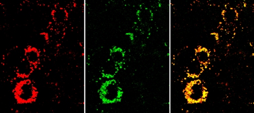
 The yellow nanosensor signal in the overlay image (right) shows that the cells are active. If they were unhealthy, they would appear much redder. Center: the indicator dye signal. Left: the reference dye signal. © Fraunhofer EMFT
The sensor nanoparticles synthesized by the research team can be used to study the impact of chemical compounds on live cells. Healthy cells stack adenosine triphosphate (ATP) for energy. If metabolic activity is at a high level in the cells, they produce more ATP. Thus, the damaged cells can only generate lower amount of ATP. According to Schmidt, the innovative nanosensor is capable of determining the amount of ATP in order to assess the health status of the cells, which in turn helps determining the damages caused by medical chemicals and compounds on the cells.
The research team used a green dye as an indicator to detect the presence of ATP and a red dye as a reference, which retains its color. The team then delivered the nanoparticles into live cells and studied them using a fluorescence microscope. If the overlay image is more yellowish in color, it indicates that the cells are active and healthy. On the other hand, if the image is more reddish in color, then they are damaged cells. The nanosensors can even be used to determine the anti-cancer activity of newly synthesized chemotherapy agents, said Schmidt.
These nanoparticles are non-toxic to cells and can easily travel via cell membranes. They can even be led to the targeted areas easily. The research team refined its work to fabricate nanosensors that are capable of identifying oxygen and toxic amine levels to detect packaged meat quality and its conditions for consumption.