A team of researchers from the University of California (UC) Berkeley and the Lawrence Berkeley National Laboratory (Berkeley Lab) has devised a technique that allows the encapsulation of nanocrystal liquids between graphene layers, to take images of chemical reactions occurring in liquid media at an atomic-scale resolution using an electron microscope.
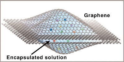
 In the graphene liquid cell, opposing graphene sheets form a sealed liquid nanoscale reaction chamber that is transparent to an electron microscope beam. The cell allows nanocrystal growth, dynamics and coalescence to be captured in real time at atomic resolution via a transmission electron microscope. (Credit: Lawrence Berkeley National Laboratory)
This new technique paves the way to directly observe biological, chemical and physical phenomena occurring in liquids by making movies at the atomic-scale resolution. Jungwon Park, one of the researchers, stated that the new graphene liquid cell allowed the researchers to encapsulate a trace quantity of liquid sample under a condition of high vacuum to capture real-time movies of the platinum nanocrystal growth. The high resolution and contrast is a result of realistic sample conditions provided by the liquid cell that mainly owes to the ultrathin thickness and chemical inertness of graphene.
Liquid cells with a viewing window are used to hermetically seal liquid samples for performing the electron microscope study. So far, these cells have been equipped with viewing windows made of silicon oxide or silicon nitride. However, the high thickness of these silicon-based cell windows prevents the electron penetration, thus limiting resolution. Moreover, these windows disturb the innate state of the liquid or specimen in the liquid.
Park explained that graphene is strong, highly impermeable, and chemically inert and allows the electron beam to traverse, thus protecting the sample present in the liquid cell from an electron microscope’s high-energy beam. To create the graphene liquid cell, a platinum growth solution was pipetted to get it encapsulated between a pair of laminated graphene layers suspended on the grid holes of a traditional transmission electron microscope (TEM). Kwanpyo Kim, one of the researchers, explained that the strong van derWaals interaction between the two graphene layers enabled them to encapsulate liquid droplets of sizes 6-200 nm.
The research team tested the graphene liquid cells using the Transmission Electron Aberration-corrected Microscope I (TEAM I) at the Berkeley Lab’s National Center for Electron Microscopy. With the help of graphene liquid cells and TEAM I, the team made first ever real-time movies of platinum nanocrystal growth in liquid at an unprecedented resolution with least sample perturbation. The team’s next step is to explore the growth of other nanoparticles. These graphene liquid cells can also be used to study biomaterials like proteins and DNA.