Oct 10 2012
The fracture of materials is a very important issue for both structural and functional materials. Crack-tip behavior is among the most basic problems in fracture mechanics. Many theoretical and experimental investigations have been carried out to understand the effect of crack-tip deformation fields.
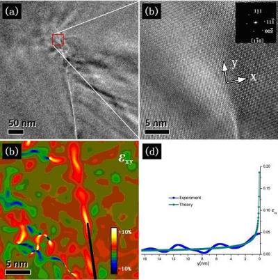
 This shows the: (a) TEM bright-field image of a micro-crack in single-crystal silicon; (b) HRTEM image of a crack-tip (boxed area in figure a); (c) shear strain field of a crack-tip; and (d) strain-distance curves on the crack plane. Credit: ©Science China Press
However, direct nanoscale measurement of strain fields around the crack-tip has not yet been achieved, despite many years of research. Professors Zhao ChunWang and Xing YongMing from Inner Mongolia University of Technology set out to tackle this problem. They adopted a combination of high-resolution transmission electron microscopy (HRTEM) and geometric phase analysis (GPA) and mapped the near atomic-scale strain fields of a crack-tip. Their work, entitled "Quantitative analysis of nanoscale deformation fields of a crack-tip in single-crystal silicon", was published in SCIENCE CHINA Physics, Mechanics & Astronomy. 2012, Vol. 55(6).
Although the fracture of materials is part of our everyday experience, the process is not well understood. The effects of fracture of materials are obvious on macroscopic scales, but the dynamics of fracture that lead to such failures are governed entirely by the material's behavior at the smallest scale. Deformation fields of a crack-tip were examined using HRTEM, which is becoming an increasingly powerful tool for measuring deformation fields at the nanoscale because of the development of quantitative image analysis methods. One such technique is GPA, which has been applied to a wide variety of systems, such as dislocations, quantum dots, nanowires, nanoparticles, heterostructures, low-angle grain boundaries, etc. The current work presented an HRTEM investigation of a mode II crack in single-crystal silicon. First, a micro-crack in single-crystal silicon was found using TEM under low magnification (Figure a). Second, the crack-tip was located and a higher magnification HRTEM image obtained (Figure b). Third, the GPA method was employed to map the strain fields of the crack-tip area from the HRTEM image (Figure c). Because the HRTEM images are at near atomic-scale, the strain map is also at near atomic-scale. The measurements showed that the strain at the crack tip was pure shear strain, and thus the micro-crack was identified as a mode II crack. Finally, the experimental strain values on the crack plane were extracted and compared with theoretical predictions from linear elastic fracture mechanics (Figure d). The comparison shows that the experimental results on the crack plane agree with the theoretical predictions up to 1 nm from the crack-tip.
Until now, experimental research on micro-cracks has mainly focused on initiation, extension and morphology at a wide variety of scales. Professor Zhao ChunWang and his group have pioneered quantitative analysis of nanoscale deformation fields of crack-tips and developed an effective method to analyze the deformation of crack-tips at a very small scale. With further development of HRTEM, measurement and analysis of deformation fields at the atomic scale will become possible. Atomic bonding and atom movement ahead of the crack-tip may be also analyzed quantitatively.
This is an important development in the recent history of fracture mechanics. The researchers suggest that the next steps in their work are to examine more materials and also to attempt to measure the deformation fields of a dynamic crack-tip. These future efforts will be an important boost to fracture mechanics and materials physics.