Feb 8 2013
In the world of microscopy, this advance is almost comparable to the leap from photography to live television. Two young EPFL researchers, Yann Cotte and Fatih Toy, have designed a device that combines holographic microscopy and computational image processing to observe living biological tissues at the nanoscale. Their research is being done under the supervision of Christian Depeursinge, head of the Microvision and Microdiagnostics Group in EPFL's School of Engineering.
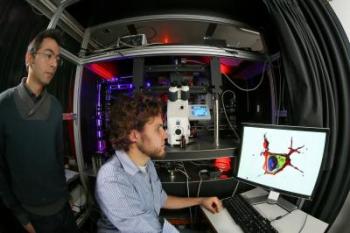
 Yann Cotte (left) and Fatih Toy are using their setup -- a holographic microscope at the back and visualization computer -- to get accurate 3-D models of live cells. Credit: Alain Herzog / EPFL
Using their setup, three-dimensional images of living cells can be obtained in just a few minutes – instantaneous operation is still in the works – at an incredibly precise resolution of less than 100 nanometers, 1000 times smaller than the diameter of a human hair. And because they're able to do this without using contrast dyes or fluorescents, the experimental results don't run the risk of being distorted by the presence of foreign substances.
Being able to capture a living cell from every angle like this lays the groundwork for a whole new field of investigation. "We can observe in real time the reaction of a cell that is subjected to any kind of stimulus," explains Cotte. "This opens up all kinds of new opportunities, such as studying the effects of pharmaceutical substances at the scale of the individual cell, for example."
Watching a neuron grow
This month in Nature Photonics the researchers demonstrate the potential of their method by developing, image by image, the film of a growing neuron and the birth of a synapse, caught over the course of an hour at a rate of one image per minute. This work, which was carried out in collaboration with the Neuroenergetics and cellular dynamics laboratory in EPFL's Brain Mind Institute, directed by Pierre Magistretti, earned them an editorial in the prestigious journal. "Because we used a low-intensity laser, the influence of the light or heat on the cell is minimal," continues Cotte. "Our technique thus allows us to observe a cell while still keeping it alive for a long period of time."
As the laser scans the sample, numerous images extracted by holography are captured by a digital camera, assembled by a computer and "deconvoluted" in order to eliminate noise. To develop their algorithm, the young scientists designed and built a "calibration" system in the school's clean rooms (CMI) using a thin layer of aluminum that they pierced with 70nm-diameter "nanoholes" spaced 70nm apart.
Finally, the assembled three-dimensional image of the cell, that looks as focused as a drawing in an encyclopedia, can be virtually "sliced" to expose its internal elements, such as the nucleus, genetic material and organelles.
Toy and Cotte, who have already obtained an EPFL Innogrant, have no intention of calling a halt to their research after such a promising beginning. In a company that's in the process of being created and in collaboration with the startup Lyncée SA, they hope to develop a system that could deliver these kinds of observations in vivo, without the need for removing tissue, using portable devices. In parallel, they will continue to design laboratory material based on these principles. Even before its official launch, the start-up they're creating has plenty of work to do - and plenty of ambition, as well.