Oct 31 2013
By the time people began to look for viable routes on the ocean with the help of a compass needle in the 12th century, magnetic navigational aids had already long been in use by other living creatures. Migratory birds orient themselves with respect to the Earth’s magnetic field, but some unicellular organisms, called magnetotactic bacteria, do as well. They carry within them a chain of nanoparticles of magnetite, a magnetic mineral that functions as an internal compass.
Details of how the microorganisms form the mineral magnetite from oxides of iron are being presented in two current publications by scientists of the Max Planck Institute of Colloids and Interfaces, Potsdam-Golm, together with colleagues from France and the USA. According to the articles, the bacteria create magnetite nanoparticles through an intermediate step that is similar to that in higher life forms; however, they use a different protein, MamP, to control the oxidation of the iron.
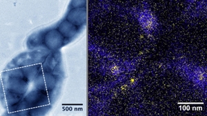
 A bacterium builds its compass needle: Magnetite particles appear clearly as dark gray structures in the transmission electron microscope image (left) that have formed within ten minutes of the unicellular organism having come into contact with a ferrous nutrient solution. An analysis of the elements in the marked section shows that iron (yellow) and phosphorus (blue) are simultaneously taken up (right).© MPI of Colloids and Interfaces / J. Baumgartner
When magnetotactic bacteria follow their internal compass, they are not seeking the proper route from north to south, but instead are seeking the bottom of oceans, rivers, or other bodies of water. This is because the microorganisms find the ideal oxygen-poor conditions for their nutrients a few millimetres below the boundary between the water and bottom sediments. They follow the lines of the Earth’s magnetic field there, which do not run parallel to the Earth's surface when far from the equator, but instead angle downward toward the surface. The bacteria orient themselves with respect to the magnetic field with the help of magnetosomes: nanoparticles of magnetite encapsulated in membranes that line up in chains along the cell axes.
Two international teams, both including scientists from the Max Planck Institute for Colloids and Interfaces, have now investigated more closely how the tiny iron oxide particles form. “Magnetotactic bacteria are excellent subjects for studying magnetic biomineralisation”, says Damien Faivre, Leader of the Molecular Biomimetic and Magnetic Biomineralisation Research Group at the Max Planck Institute in Potsdam. “This is because their genomes are already decoded and there are enough investigative techniques with which we can genetically alter them.”
The way bacteria control the biomineralisation offers a great model for materials science
In general, magnetite crystals consist of iron(II,III) oxide (Fe3O4) containing two different species of iron. The shape of the nanoparticle and thus the magnetosomes varies among different species of bacteria, though one species of bacteria always forms them with great precision in the same shape and size. The microbes are apparently able to control the biosynthesis of the nanoparticles in a unique manner, which has awakened the interest of not just biologists. “If we develop a better understanding of the underlying principles, new approaches and methods of producing magnetite nanoparticles in the future will certainly open up”, according to Damien Faivre. “If materials scientists were able to control the properties of synthetic magnetite particles as precisely as bacteria do, then new applications would be conceivable for the particles, such as contrast media for magnetic resonance tomography.”
In one of the recently published studies, the scientists chemically characterised how the magnetotactic bacteria formed magnetite using X-ray absorption spectroscopy at cryogenic temperatures and transmission electron microscopy. The unicellular organisms first create completely disordered ferric hydroxide rich in phosphate. This material resembles ferritins, i.e. a protein complex occurring in animals, plants and bacteria that typically stores iron. The magnetite particles for the magnetosomes are subsequently formed from nanoparticles of ferric oxyhydroxides or oxides.
“The astounding thing is that this transformation to magnetite is very similar to and resembles how the mineralisation in higher organisms works”, says Jens Baumgartner, one of the participating scientists of the Max Planck Institute for Colloids and Interfaces. Pigeons also probably form magnetite using the same mechanism in order to deposit it in their beaks as a navigational aid. Since iron phosphates are present, it suggests that the biomineralisation of magnetite in bacteria and higher life forms proceeds similarly, even though these life forms are widely separated from each other from an evolutionary point of view.
Bacteria control the oxidation of iron through the MamP protein
However, the bacteria are not just able to precisely control the shape and size of the magnetite particles, but the chemical composition of the particles as well. They create the exact chemical conditions for the oxides of the two different iron ions, i.e. the doubly charged iron(II) (Fe(II)) and the triple-charged iron(III) (Fe(III)), to be formed in exactly the right proportions. As the second study now shows, in which the scientists in Potsdam were also involved, a protein named MamP plays the critical role. This protein was found exclusively in magnetotactic bacteria and resembles cytochromes.
Cytochrome transfers electrons in redox reactions during cellular respiration and other biochemical processes. The researchers have now established that MamP oxidises Fe(II) to Fe(III). The bacteria therefore only need iron(II) in order to create the magnetite particles. By having genetically altered the protein, the scientists also identified the structural elements important for the iron oxidation reaction, among other results. These subunits of the protein are labelled magnetochromes.
“Until now, it was not known whether the bacteria started with iron(II) or iron(III) when forming magnetite”, explains Damien Faivre. “Our study has now resolved this question.” The answer also corresponds to what one would expect from the environment of the bacteria: in the oxygen-poor layer where the microbes live, iron is present in the more weakly oxidised form, i.e. that of iron(II).