Mar 31 2014
Umeå University in Sweden will be a national resource for advanced electron microscopy thanks to a contribution of SEK 57 million. Scientists across the country will be able to use the techniques for research, including detailed studies of microorganisms that cause infectious diseases.
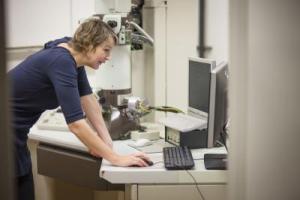
 This is a photo of Dr. Linda Sandblad at the Umeå Core Facility Electron Microscopy. Credit: Mattias Pettersson
"Sweden needs a national platform with a state-of-the-art electron microscopy facility to meet the future needs of visualization in medical, chemical and biological research. Umeå will now become a central node in a national network of research groups working together to improve the methods. This is not only a strength for our researchers, but also for the university as a whole," says Lena Gustafsson, Vice-Chancellor at Umeå University.
The Knut and Alice Wallenberg Foundation is investing SEK 37 million to develop the platform. The main applicants are Bernt Eric Uhlin and Linda Sandblad, Umeå Centre for Microbial Research (UCMR), who along with colleagues at Umeå University, Karolinska Institutet, Linköping University, Lund University and the Swedish University of Agricultural Sciences have developed plans for a national platform in Umeå.
Umeå University is also supporting the venture with another SEK 15 million. Moreover, the Kempe Foundation is investing SEK 5 million, which allows the acquisition of the most advanced and powerful equipment for electron microscopy.
"Thanks to the funding we can make a major upgrade of our existing technology, we will, for example, be able to create 3D electron microscopy images with the best possible resolution and visualize molecular details of bacteria and other cells," says Linda Sandblad, group leader at UCMR and director of Umeå Core Facility Electron Microscopy (UCEM), located in the Umeå University's Chemical Biological Centre (KBC), where the new equipment will be included.
"This new grant further strengthens our commitment to scientific infrastructure within the KBC, "said Per Gardeström professor and scientific coordinator for KBC."
"It shows again that the research environment at Umeå Unversity which is based on collaboration over faculty and university boundaries is very successful and by sharing of internationally competitive equipment, we will hopefully also in the future attract top researchers at Umeå."
The funding will be used to enhance two different techniques. The first technique is called cryo-electron microscopy, which makes it possible to study the smallest building blocks of life – proteins and lipid membranes – such as they occur in their natural water-soluble environment. The resolution is at atomic level. With the second technique, electron tomography, it is possible to visualise and analyse a sample in three dimensions. A sample with, for example, bacterial cytoskeleton can be rotated in the electron microscope and visualised from different angles.
"We study how microorganisms cause infectious diseases and how all living organisms function biochemically at molecular level," says Bernt Eric Uhlin, professor of medical microbiology and director of MIMS (Laboratory for Molecular Infection Medicine Sweden), which is the Swedish node in the international cooperation of molecular medical research with support from the Swedish Research Council.
"By visualising protein complexes and membrane structures, we learn how they are structured and what mechanisms and functions they fulfil in the cell."
Two new electron microscopes and tomography equipment will be purchased to the national platform thanks to new funding. The techniques will be very important to a large number of research projects conducted at Swedish universities. It will also further strengthen the international research centres in Umeå: UCMR, MIMS and Umeå Plant Science Centre (UPSC) with the Berzelius Centre for Forest Biotechnology.