Mar 13 2015
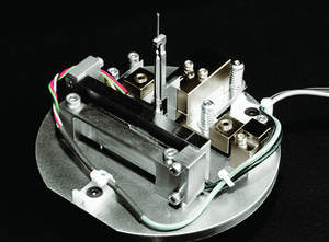
ZEISS announces the new Ultra Load Stage for ZEISS Xradia Ultra 3D X-ray microscopes (XRM). Xradia Ultra Load Stage uniquely enables in situ nanomechanical testing -- compression, tension, indentation -- with non-destructive 3D tomographic imaging. For the first time, researchers will be able to image the evolution of structure in 3D under load down to 50 nm resolution. This new capability applies to a wide range of interests, covering both engineered and natural materials.
 Xradia Ultra Load Stage for ZEISS Xradia Ultra 3D X-ray Microscopes: compression, tension and indentation studies at the nanoscale
Of particular interest to materials scientists, Xradia Ultra Load Stage explores a new critical length scale for in situ materials characterization, enabling researchers to observe internal features such as nanoscale cracks and voids that ultimately lead to deformation and failure at the macroscale. This capability supports "materials by design," a concept that enables the development of unique new materials for function-specific applicability, such as lighter, stronger fiber composites for airplane wings; more durable concrete for building materials; and biomimetic materials reproducing the advanced mechanical properties of many natural structures.
The University of Manchester in Manchester, UK has performed 3D in situ imaging of crack growth using Xradia Ultra Load Stage in nanoindentation mode to understand how cracks grow in dentin, the nano-composite that forms the bulk of teeth. Philip Withers, Professor of Materials Science and Director of the Manchester Henry Moseley X-ray Imaging Facility, says, "In situ nanomechanical testing in the Xradia Ultra X-ray microscope has enabled us to link the nanoscale 3D structure of a material directly to its performance. For the first time we can now undertake time-lapse 3D imaging with phase contrast to follow the nucleation and growth of defects and cracks in both natural and synthetic nanostructured materials."
Los Alamos National Laboratory has already studied a variety of materials with the instrument, including glass microspheres, foams, metal alloys, and single microcrystals. A team led by Brian M. Patterson uses the stage to better understand damage initiation and growth in materials, gaining insight into how processing affects mechanical response.
Dr. Kevin Fahey, chief materials scientist and head of Carl Zeiss X-ray Microscopy, notes that the development of Ultra Load Stage was undertaken in a continuing effort to enable the company's customers to advance science with unique new capabilities. "ZEISS Xradia Ultra X-ray microscopes provide the highest resolution available in non-destructive 3D X-ray imaging outside a synchrotron facility. Now, our customers are able to bridge the lengthscale/force gap between the micron scale and in situ SEM/TEM, visualizing and quantifying 3D nanostructures under load. This is a completely unique capability that offers new opportunities to connect small scale evolution processes with those observed in micron scale XRM and bulk material testing."
Ultra Load Stage satisfies a void in the current availability of mechanical testing and imaging approaches, offering new capabilities to observe internal processes at nanoscale resolution. Until now, electron microscopy techniques provided resolution down to the nanometer range but are limited to surface imaging (SEM) or required extremely thin samples whose mechanical behavior will be strongly affected by surface effects (TEM).
Ultra Load Stage comprises a piezomechanical actuator with closed loop position control, a strain gauge force sensor and anvil sets that can be configured for three different operating modes, compression, tension, and indentation. It is available immediately.
Visit www.zeiss.com/xrm for more information about ZEISS Xradia 3D X-ray microscopes. To download information about Xradia Ultra Load Stage, visit http://info.xradia.com/Xradia-Ultra-Load-Stage.html.
ZEISS
The Carl Zeiss Group is an international leader in the fields of optics and optoelectronics. In fiscal year 2011/12 the company's approximately 24,000 employees generated revenue of nearly 4.2 billion euros. In the markets for Industrial Solutions, Research Solutions, Medical Technology and Consumer Optics, ZEISS has contributed to technological progress for more than 160 years and enhances the quality of life of many people around the globe. The Carl Zeiss Group develops and produces planetariums, eyeglass lenses, camera and cine lenses and binoculars as well as solutions for biomedical research, medical technology and the semiconductor, automotive and mechanical engineering industries. ZEISS is present in over 40 countries around the globe with about 40 production facilities, over 50 sales and service locations and service locations and approximately 20 research and development sites. Carl Zeiss AG is fully owned by the Carl Zeiss Stiftung (Carl Zeiss Foundation). Founded in 1846 in Jena, the company is headquartered in Oberkochen, Germany.
Microscopy
The Microscopy business group at ZEISS is the world's only manufacturer of light, X-ray and electron microscopes. The company's extensive portfolio enables research and routine applications in the life and materials sciences. The product range includes light and laser scanning microscopes, X-ray microscopes, electron and ion microscopes and spectrometer modules. Users are supported for software for system control, image capture and editing. The Microscopy business group has sales companies in 33 countries. Application and service specialists support customers around the globe in demo centers and on site. The business group is headquartered in Jena, Germany. Additional production and development sites are in Oberkochen, Göttingen and Munich, as well as in Cambridge in the UK and Peabody, MA and Pleasanton, CA in the USA. The company has around 2,800 employees and generates revenue of 650 million euros.