Feb 11 2016
Photographers capture high-speed motion using cameras with a split-second shutter speed feature. Similar to these photographers, scientists examine very small materials using specific instruments, which are capable of seeing changes that occur in the blink of an eye.
A team of researchers from all over the world, headed by X-ray scientist Christoph Bostedt of the U.S. Department of Energy's (DOE's) Argonne National Laboratory, and Tais Gorkhover of DOE's SLAC National Accelerator Laboratory, used two special lasers to examine the dynamics of a xenon sample when heated to a plasma.
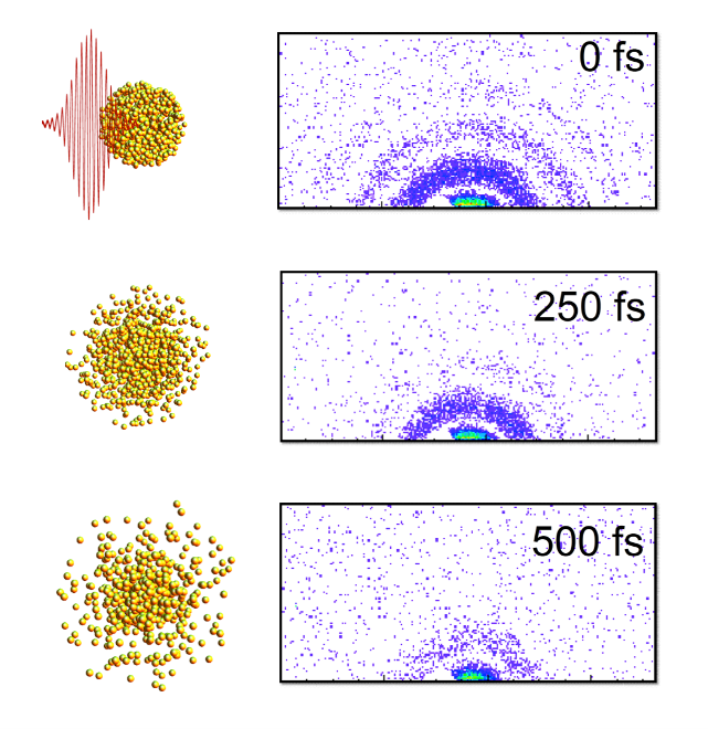
 Here are “stills” from an X-ray “movie" of an exploding nanoparticle. The nanoparticle is superheated with an intense optical pulse and subsequently explodes (left). A series of ultrafast X-ray diffraction images (right) maps the process and contains information how the explosion starts with surface softening and proceeds from the outside in.
Gorkhover and Bostedt used the Linac Coherent Light Source (LCLS) at SLAC to observe the sample steps, timed to about a hundred femtoseconds, where one femtosecond refers to one millionth of a billionth of a second. The particles moving very fast in the gas phase were appeared frozen, due to the short exposure time of every single image.
The advantage of a machine like the LCLS is that it gives us the equivalent of high-speed flash photography as opposed to a pinhole camera.
Christoph Bostedt, X-Ray Scientist, U.S. Department of Energy's Argonne National Laboratory
The team heated the sample cluster using an optical laser, and examined the dynamics of the cluster with the help of an X-ray laser. The heating of the cluster with the laser resulted in the emission of electrons from atoms by the photons. The electrons continued to remain loosely bound to the cluster.
The team measured how the nanoparticles changed when exposed to rough environments by imaging exploding nanoparticles.
Ultimately, we want to understand how the energy from the light affects the system. There are really no other techniques that give us this good a resolution in both time and space simultaneously. Other methods require us to take averages over many different 'exposures,' which can obscure relevant details. Additionally, techniques like electron microscopy involve a substrate material that can interfere with the behavior of the sample.
Tais Gorkhover, U.S. Department of Energy's SLAC National Accelerator Laboratory
Bostedt stated that the research could influence the study of aerosols in combustion or in the environment, because the dual-laser "pump and probe" model could be used to analyze materials existing in the gas phase.
Although our material goes from solid to plasma very quickly, there are other types of materials you could study with this or a similar technique.
Christoph Bostedt, X-Ray Scientist, U.S. Department of Energy's Argonne National Laboratory
The February 2016 issue of Nature Photonics published an article on "Femtosecond and nanometer visualization of structural dynamics in superheated nanoparticles".
The research was financially supported by DOE's Office of Science. Tais Gorkhover received a Peter Paul Ewald Fellowship from the Volkswagen Foundation.