Mar 1 2016
The infection mechanisms of coronaviruses can now be examined in detail by using supercomputing processes and high-resolution cryo-electron microscopy. These viruses are known for attacking the respiratory tract of animals and humans.
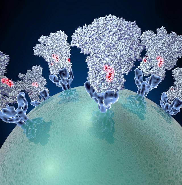
 Coronaviruses -- the agents behind outbreaks of new kinds of pneumonia -- employ molecular tactics to infect cells. Veesler Lab/University of Washington
A team of researchers from the University of Utrecht, the University of Utrecht, and the University of Washington (UW) has developed an atomic model of a coronavirus spike protein that encourages the cellular entry of coronaviruses. A number of ideas for particular vaccine strategies have been developed through an analysis of this model. The findings are featured in a new UW Medicine-led study published in the journal Nature. The project was led by David Veesler, UW assistant professor of biochemistry.
Veesler stated that these viruses containing spiked crowns are globally accountable for a third of cold-like, mild symptoms, and atypical pneumonia. However, in 2002 the most dangerous forms of coronaviruses appeared in the form of severe acute respiratory syndrome coronavirus (SARS-CoV), and in 2012 coronaviruses further existed in the form of Middle East respiratory syndrome coronavirus (MERS-CoV), thus resulting in 10% to 37% of mortality rates.
The deadly pneumonia epidemic highlighted that coronaviruses are capable of being transferred from different animals to humans. At present, a total of only six coronaviruses are believed to have infected people, but animals are naturally affected by a huge number of coronaviruses. The recent epidemic outbreaks are the result of coronavirus conquering the species barrier. This highlights the possibility of the emergence of new coronavirus with pandemic potential. Approved antiviral treatments and vaccines are still not available for MERS-CoV or SARS-CoV.
A transmembrane spike glycoprotein helps in mediating the capability of coronaviruses to fasten and then enter particular cells. Trimers are structures collected from three protein units that are identical. The team examined the structure responsible for attaching to and combining with a living cell’s membrane. The spike establishes the kinds of animals and cells in their bodies that every single coronavirus is capable of infecting.
Veesler and his team displayed the architecture of a mouse coronavirus spike glycoprotein trimer by using a recently developed single particle cryo-electron microscopy and supercomputing analysis. The team detected that the resolution is 4 angstroms, which is a measurement unit expressing the size of atoms and the distances between each atom that is equal to one-tenth of a nanometer.
The structure is maintained in its pre-fusion state, and then undergoes major rearrangements to trigger fusion of the viral and host membranes and initiate infection.
David Veesler, UW assistant professor of biochemistry.
The coronavirus fusion machinery resembles to the blend of proteins present in the paramyxoviruses, which is another virus family including syncytial virus and the viruses responsible for mumps and measles. The syncytial virus is responsible for causing wheezing in children as well as infant hospitalizations. This resemblance highlights that the fusion proteins of coronavirus and paramyxovirus are capable of using very similar mechanisms for encouraging viral entry and sharing a familiar evolutionary origin.
A comparative analysis was conducted for crystal structures of the spike protein present in mouse and human coronaviruses. The results provide hints on how the molecular structure of the protein domains might impact which particular animal species the virus is capable of infecting.
The structure for attainable targets for anti-viral therapies and vaccine design was also examined by the researchers. The team discovered that the exterior edge of the coronavirus spike trimer’ has a blend of fusion peptide, which is a chain of amino acids, involved in the entry of viruses into host cells. Vaccine strategies that are capable of neutralizing a wide range of these viruses are suggested through the effortless accessibility of this peptide and its predicted resemblance among several coronaviruses.
Our studies revealed a weakness in this family of viruses that may be an ideal target for neutralizing coronaviruses
David Veesler, UW assistant professor of biochemistry.
The researchers highlighted that there may be a way to obtain broadly neutralizing antibodies that recognize this peripheral peptide. Neutralizing antibodies help protect infections by arresting a mechanism in a pathogen. Broadly neutralizing antibodies are believed to be effective against a number of pathogen strains, in this case coronaviruses. The fusion peptide’s physical structure stimulates ideas for protein designs that would disable it.
"Small molecules or protein scaffolds might eventually be designed to bind to this site," Veesler said, "to hinder insertion of the fusion peptide into the host cell membrane and to prevent it from undergoing changes conducive to fusion with the host cell. We hope that this might be the case, but much more work needs to be done to see if it is possible."
The Letter to Nature contains a description of the coronavirus spike protein structure, which is expected to be very similar to other coronavirus spike proteins.
"Therefore, the structure we analyzed in the mouse coronavirus is likely to be representative of the architecture of other coronavirus spike proteins such as those of MERS-CoV and SARS-CoV," the researchers observed.
The researchers summarized their paper, "Our results now provide a framework to understand coronavirus entry and suggest ways for preventing or treating future coronavirus outbreaks."
"Such strategies," Veesler said, "would be applicable to several existing coronaviruses and to emerging future strains of coronavirus that conserve this same structure for entering cells."