Jun 21 2017
A Franco-Dutch International team including Scientists from the laboratories of Condensed Matter Physics and Hydrodynamics at Paris-Saclay University and the Van’t Hoff Institute for Molecular Sciences at the University of Amsterdam has developed an innovative technique for accurately determining, in real time, the fluid flow in capillary networks.
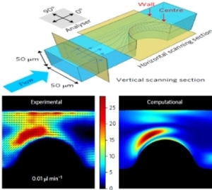
 Schematic illustration of the microfluidic channel (above) and the experimental result obtained using the nanorods (below, left) which is in good agreement with the computational result (below, right). Credit: University of Amsterdam
The proof-of-concept for the research has been reported in the latest issue of the journal Nature Nanotechnology.
Professor of Spectroscopy and Photonic Materials Fred Brouwer and Research Technician Michiel Hilbers, both from HIMS, performed confocal imaging and single-particle measurements of the nanorods. LaserLab Europe supported the team.
The analysis of fluid flow in capillary networks proves to be advantageous in various fields. For instance, determining the circulation of blood in arteries is a significant facet of the analysis of the formation of plaque in atherosclerosis. While hydrodynamic simulations can produce significant information, experimental investigations are necessary for eventual confirmation.
Yet, it is highly difficult to characterize fluid flows of the order of a few hundred nanometers. The prevalent method of particle imaging velocimetry (PIV) that tracks the displacements of fluorescent microspheres cannot be applied for locally observing dynamic systems in real time. Moreover, considering velocity gradients (shear) that are common to capillary networks, PIV exhibits restricted spatial resolution and weak signal-to-noise ratios.
At present the French-Dutch International research team has described the application of nanorods instead of spheres, in the Nature Nanotechnology paper. They have demonstrated that when the collective orientation of the nanorods is instantly identified in a small focal volume, the local shear rate can be directly measured and quickly scanned. As a proof-of-principle, they showed that tomographic mapping of the shear distribution can be performed in a microfluidic model system by means of scanning confocal microscopy.
The team developed nanorods formed of lanthanum phosphate, or LaPO4, crystals that were doped by using luminescent europium, or Eu3+, ions. Like logs of trees floating on a river these nanorods, 10 nm in diameter and 200 nm in length, gradually orient themselves along the direction of flow. The spatial orientation of the Eu3+ ions could be traced through their emission spectrum due to the strongly polarized light emission characteristics of the ions. Consequently, fluid flow in a small-sized microfluidic channel was feasibly analyzed in real time with unmatched resolution.
This work opens promising perspectives for the fundamental understanding of phenomena related to the flow of a fluid in complex channels. In the future, such orientation probes can be applied to the field of biology in order to understand in-situ complex mechanisms involved in the dynamics of orientation of bio-macromolecules to account for their characteristics and functions.