Mar 8 2019
Using wonder material graphene, scientists at the University of Minnesota College of Science and Engineering have created an innovative device that lays the foundation for developing ultrasensitive biosensors for the detection of a host of diseases at the molecular level, with almost unprecedented efficiency.
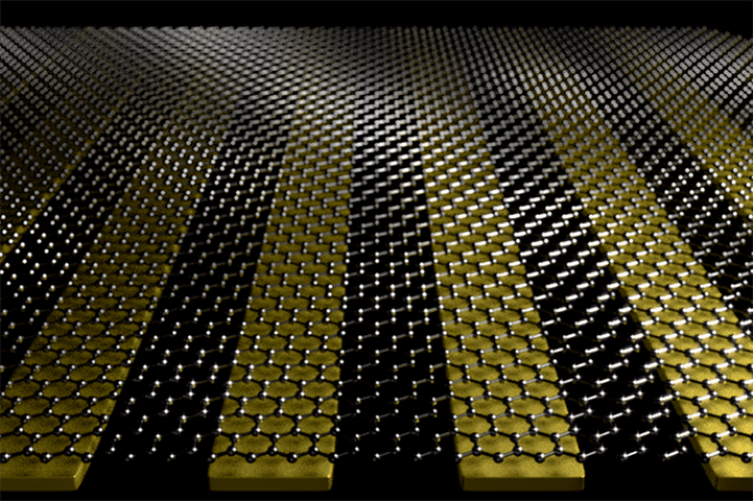
 University of Minnesota researchers combined graphene with nano-sized metal ribbons of gold to create an ultrasensitive biosensor that could help detect a variety of diseases in humans and animals. (Image credit: Oh Group, University of Minnesota)
The use of ultrasensitive biosensors for studying the structures of proteins can considerably enhance the depth of diagnosis for many types of diseases, extending to both animals and humans. These disorders include mad cow disease, Chronic Wasting Disease, and Alzheimer’s disease, all associated with protein misfolding. Such kinds of biosensors could also give way to better technologies for producing novel pharmaceutical compounds.
The study has been reported in a peer-reviewed scientific journal Nature Nanotechnology, published by Nature Publishing Group.
In order to detect and treat many diseases we need to detect protein molecules at very small amounts and understand their structure. Currently, there are many technical challenges with that process. We hope that our device using graphene and a unique manufacturing process will provide the fundamental research that can help overcome those challenges.
Sang-Hyun Oh, Study Lead Researcher and Professor, Department of Electrical and Computer Engineering, University of Minnesota.
Graphene is a unique material composed of one layer of carbon atoms. Discovered more than 10 years ago, the material has fascinated scientists with its range of incredible properties that have found applications in a variety of innovative applications, including the development of improved sensors for detecting various diseases.
While considerable efforts have been made to enhance biosensors through graphene, the challenge continues to exist because of the material’s unique single atom thickness. This implies that when a light is shined through graphene, the latter does not interact well with the former. During the diagnosis of diseases, the absorption and conversion of light to local electric fields is important for detecting trace amounts of molecules. Earlier studies using analogous graphene nanostructures only showed a light absorption speed of less than 10%.
In the latest study, researchers from the University of Minnesota integrated nano-sized metal ribbons of gold with graphene. With the help of sticky tape as well as a high-tech nanofabrication method devised at the University of Minnesota, known as “template stripping,” the team successfully produced an extremely flat base layer surface for the graphene.
The researchers subsequently applied light energy to produce a sloshing movement of electrons within the graphene, referred to as plasmons, which can be assumed to be like waves or ripples dispersing through a “sea” of electrons. In a similar way, such waves can grow in intensity to huge “tidal waves” of local electric fields on the basis of the team’ ingenious design.
When the researchers shined a light on the one-atom-thick graphene layer device, they successfully produced a plasmon wave with near perfect efficiency of 94% light absorption into “tidal waves” of electric field. Moreover, the insertion of protein molecules between the metal ribbons and the graphene allowed the researchers to harness sufficient amounts of energy to visualize monolayers of protein molecules.
“Our computer simulations showed that this novel approach would work, but we were still a little surprised when we achieved the 94 percent light absorption in real devices,” stated Oh, who holds the Sanford P. Bordeau Chair in Electrical Engineering at the University of Minnesota. “Realizing an ideal from a computer simulation has so many challenges. Everything has to be so high quality and atomically flat. The fact that we could obtain such good agreement between theory and experiment was quite surprising and exciting.”
Apart from Oh, the research group included University of Minnesota electrical and computer engineering postdoctoral researchers In-Ho Lee (lead author) and Daehan Yoo, Professor Tony Low, as well as IBM Fellow Emeritus Dr Phaedon Avouris.
The study was mainly funded by the National Science Foundation. Additional support was provided by the Institute for Mathematics and its Applications (IMA) at the University of Minnesota. The device was developed at the Minnesota Nano Center at the University of Minnesota, and electron microscopy measurements were carried out at the University of Minnesota Characterization Facility.