A recent study published in the Chemical Engineering Journal focuses on the photoluminescence and bio-imaging applications of highly bio-compatible CsPbBr3 perovskite nanocrystals encased in a SiO2 nanoshell.

Study: Two-photon Photoluminescence and Bio-imaging Application of Monodispersed Perovskite-in-silica Nanocrystals with High Biocompatibility. Image Credit: Caleb Foster/Shutterstock.com
Two-photon fluorescent probes composed of synthetic lead material can be used effectively for bio-imaging applications such as diagnosis of cancer cells, but their low biocompatibility due to the high reactivity and cytotoxicity of Pb+2 ions significantly limit their usage in the bio-medical industry.
Importance of Two-Photon Fluorescence Imaging
Early detection and testing are the only successful methods for increasing the survival rate of cancer. Near-infrared (NIR) lasers can be concentrated on a very tiny area and have many benefits, including excellent spatial precision, high penetration, low noise illumination, and minimal oxidative stress to cells. Two-photon luminescence microscopy, employing two near-infrared (NIR) rays as an excitation signal and resulting in the production of light waves, is regarded as a highly effective technique for monitoring cell activities and detecting early cancer mutations.
As a result, the development of high-performance, biodegradable, and cost-effective two-photon fluorescent sensors is of considerable significance in the advancement of cancer therapy. Biomedical imaging using lanthanide-treated nanocrystals, aggregation-induced luminescence nanomaterials, and II-VI nanoparticles under near-infrared laser light has attracted a lot of interest in recent years.
Lead Bromide (CsPbBr3) Nanocrystals for Bio-imaging Applications
Because of their substantial optical absorption and magnified photoluminescence, synthetic lead perovskite nanocrystals such as CsPbBr3 have surfaced as promising materials for optical and electrical applications. However, due to the inherent instability and cytotoxicity of Pb+2 ions, their applicability in the bio-imaging sector is quite limited.
The coating of silica (SiO2) protects lead bromide nanocrystals from reactive species such as humidity, improves photo-stabilities, and reduces hazardous ion movement, which enhances the biocompatibility of NCs.
Limitations of Previous Methods
Previously, CsPbBr3 nanocrystals have been integrated inside the silica shells to reduce blinking while improving water resistance and light absorption. However, the nanocrystals produced with this approach are too large for bio-imaging applications. Another method uses ultraviolet light to enclose CsPbBr3 nanocrystals within 3.5nm thick SiO2 nano-shells, but UV light has poor penetration depth in human tissues. Therefore, the nanocrystals prepared under UV radiations are not suitable for the detection of cancer cells.
A Novel Technique for Producing Biocompatible CsPbBr3 Nanocrystals
In this work, the researchers developed lead bromide nanocrystals encapsulated in silica nanoshells in a straightforward one-step nucleation technique and constantly adjusted the silica shell thickness between 15 and 60 nm. The surface appearance and crystalline architecture of the nanocrystals were examined using a transmission electron microscope. The phase concentration was determined using X-ray diffraction, and the contents and oxidation states of the NCs were determined using X-ray photoelectron spectroscopy.
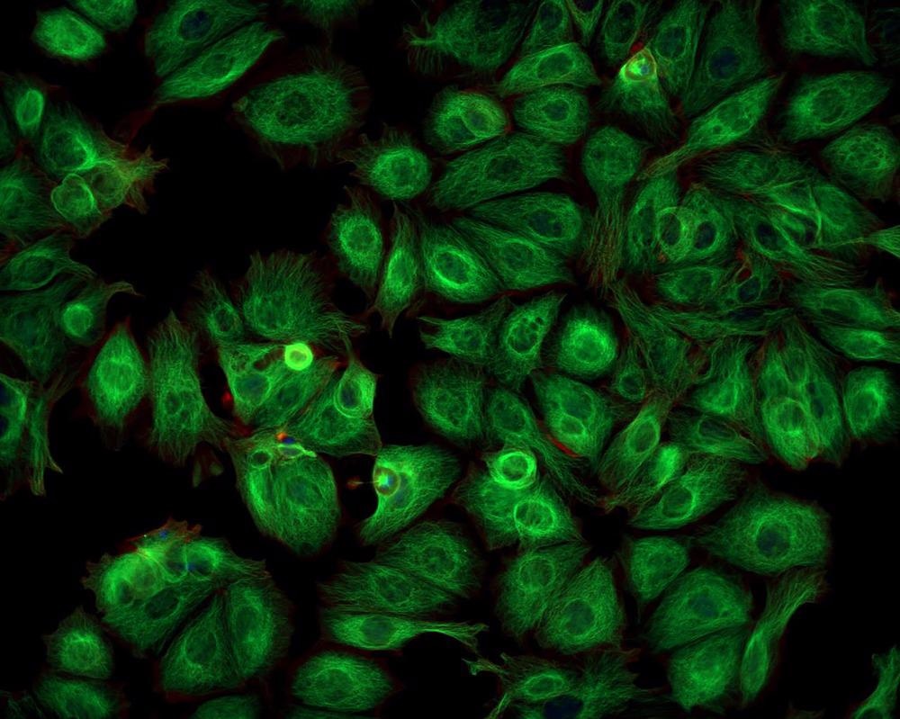
Human hepatocellular cancer cells (HepG2) were employed to test the cytocompatibility of the produced lead bromide nanocrystals. After washing with phosphate buffer salt (PBS) solution, the cultivated HepG2 cells were transferred to a fresh medium containing lead bromide nanocrystals embedded in SiO2. NCs penetrated the cancer cells via endocytosis during this process. The cells were rinsed again with PBS solution, and a tiny quantity of PBS solution was added to maintain the cells' moisture content. Finally, fluorescent images of HepG2 cancer cells were acquired using a laser scanning microscope with an 800 nm laser as the excitation source.
Important Research Findings
The researchers observed that a SiO2 shell with an appropriate thickness of 30 nm inhibited nanocrystal clumping and non-radiative mixing of charged particles. The produced nanocrystals exhibited high two-photon adsorption and generated green light peaks at 526 nm when excited by a near-infrared laser at 800 nm. The average decay life of the lead bromide nanocrystals under two-photon excitation was close to that of one-photon illumination, which is extremely useful for biomedical applications.
After 24 hours of treatment and storage with lead bromide nanocrystals embedded in silica nanoshells, the survival rate of human cancerous cells (HepG2) was determined to be 96.21 percent, confirming the NCs' exceptional cytocompatibility. The cumulative emission intensity of the produced CsPbBr3@SiO2 nanocrystals was at least five times that of the control group of nanocrystals.
In short, this research highlighted a one-step, straightforward method for the fabrication of biocompatible lead bromide nanocrystals with useful applications in the field of bio-imaging, particularly cancer cell diagnosis.
Continue reading: An Overview of Gold Thin Films: From Sensors to Cell Culture.
Reference
Zhong, C.Y. et al. (2021). Two-photon photoluminescence and bio-imaging application of monodispersed perovskite-in-silica nanocrystals with high biocompatibility. Chemical Engineering Journal. Available at: https://www.sciencedirect.com/science/article/abs/pii/S1385894721056849
Disclaimer: The views expressed here are those of the author expressed in their private capacity and do not necessarily represent the views of AZoM.com Limited T/A AZoNetwork the owner and operator of this website. This disclaimer forms part of the Terms and conditions of use of this website.