A team of researchers recently published a paper in the MDPI journal Materials that reviewed recent atomic force microscopy (AFM) investigations of halloysite nanotube (HNT) nanocomposites and HNTs to determine the feasibility of using the AFM technique for nanomaterial characterization.

Study: AFM Characterization of Halloysite Clay Nanocomposites’ Superficial Properties: Current State-of-the-Art and Perspectives. Image Credit: Gorodenkoff/Shutterstock.com
What are HNTs?
HNTs is a new one-dimensional (1D) natural nanomaterial with rich functionality, natural availability, high mechanical strength, good biocompatibility, and tubular nanostructure. It has gained attention as a cheaper alternative to multi-walled carbon nanotubes (MWCNTs).
The inner diameter of HNTs is smaller than 100 nanometers and the HNT length varies between 0.5 and 1.5 micrometers.
The difference in the surface chemistry at the outer and inner surfaces is a unique feature of HNTs as the inner surface consists of aluminum-hydroxyl groups, while the outer surface is composed of silicon-oxygen-silicon groups. Thus, the inner and outer surfaces of HNTs can be functionalized differently by grafting organosilanes, polymers, surfactants, and nanoparticles, and alkaline etching.
Moreover, these nanotubes have a notably large cavity that is suitable for confining specific molecules. This property allows the application of HNTs in the biomedical field.
Certain drugs, such as carbohydrates, proteins, and nucleic acids can be loaded into the HNT cavity by various methods, such as tubular entrapment, absorption, and intercalation, and directly delivered to specific targets in the body to prevent the degradation of bioactive molecules.
Significance of AFM for Polymer-HNT Nanocomposites Characterization
Polymer-HNT nanocomposites possess excellent biological, thermal, and mechanical properties owing to the superior properties of HNTs. The chemical composition and nanostructured surface of polymer-HNTs nanocomposites are often characterized using high-resolution microscopy techniques.
However, AFM can be used as an effective alternative to existing techniques as it allows the characterization of nanomechanical, roughness, friction, adhesive, and topographic features of the nanocomposites under an ambient environment by raster-scanning a sample surface.
AFM is currently the most powerful and versatile tool for nanoscale characterization of a sample surface as it does not require any pre-treatment of samples for characterization, unlike other microscopy techniques.
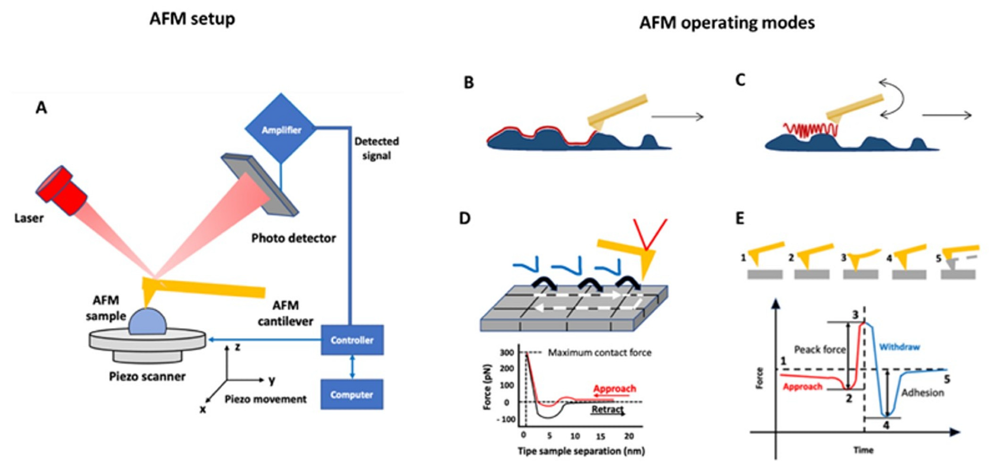
In the past few years, significant advancements have been made in AFM techniques by implementing quantitative mechanics, tip preservation, and imaging capabilities for multifrequency measurements by adding new AFM modes. For instance, tapping and contact modes are used to visualize the sample topography at high resolution and obtain information related to superficial features.

AFM Characterizations of HNT Nanocomposites
Among nano-clays, HNTs were used extensively in several application fields. This review provided an overview of the recent studies of HNT bio- and nanocomposites and HNTs performed using different modalities of the AFM technique.
Contact mode AFM was employed to characterize the morphological properties of alginate/HNTs and chitosan/HNTs bionanocomposite films. The study revealed that the addition of HNTs led to nanoscale changes on the chitosan surface, leading to an increased roughness in both alginate matrix and chitosan that resulted in greater cell adhesion capability. Thus, the findings indicated the feasibility of using these nanocomposites for tissue engineering applications.
The investigation of dried HNT-modified polyethersulfone membrane surface properties using AFM revealed that the increasing roughness on the membrane surface was correlated with the rising pH when the HNT nanofillers were loaded into polyethersulfone membranes.
Researchers used contact mode AFM to evaluate the effects of three nanoclays, including HNTs, on the Jatropha oil-enriched waterborne polyurethane matrix properties. The AFM topographies indicated that incorporating HNTs improved the properties of polymeric material.
In another study, the properties of HNT-modified polyvinyl alcohol-polyvinylpyrrolidone (PVA-PVP) composite materials were investigated using AFM. The AFM characterization of roughness parameters helped in understanding the effects of various polymerization steps, which critically influence the performance of the material in dentistry applications.
Recently, tapping mode AFM was utilized in air condition to investigate the roughness alteration of the PVA/PVP bionanocomposite films after chitosan-modified HNTs were added to the films. The AFM characterization revealed uniform dispersion of chitosan-modified HNTs on the films, which reduced the roughness of PVA/PVP films compared to the unmodified films.
Tapping mode AFM was also employed to analyze the surface roughness of the PVA/ starch (ST)/ glycerol (GL)/HNTs bionanocomposite films and HNT aspect ratios. The AFM topographies indicated that the addition of HNTs changes the roughness of the films, which in turn improves their barrier properties and makes them effective for food packaging applications.
Tapping quantitative nanoscale mechanical (QNM) mode AFM at room temperature and in air conditioning was used to characterize the HNT-doped polysulphone membrane. The AFM analysis demonstrated homogeneous HNT distribution in the matrix doped with 0.2 weight percentage HNTs, which was also confirmed by roughness quantification where the image analysis displayed a substantial reduction in root–mean–square roughness and average roughness values in 0.2 weight percentage HNT-doped membranes.
Tapping QNM mode AFM was also employed to evaluate the nanomechanical properties of doped and pristine membranes in terms of adhesion forces and elastic forces in the same study. The findings revealed a considerable increase in Young’s modulus value and the presence of aggregates on the membrane surface.
Moreover, tapping QNM mode AFM was used in a very recent study to confirm the effectiveness of a novel keratin/HNTs-based treatment of human hair. The AFM analysis confirmed that the treatment is effective for successfully protecting the hair structure.
Semi-contact mode AFM was utilized to characterize titanium dioxide (TiO2) or HNT-modified polymethylmethacrylate (PMMA). The analysis showed that the roughness value decreased substantially after nanofiller addition, specifically after HNT addition, while the stiffness increased significantly after TiO2 addition compared to HNT addition.
Significance of the Review
The review demonstrated that AFM and related techniques can be used as an effective tool to investigate the materials- and morphological-related details of HNT nanocomposites that other microscopy techniques cannot detect. HNT nano- and biocomposites characterized using AFM in recent studies displayed enhanced functional properties and mechanical strength.
These nanocomposites can be used in dentistry, cosmetics, and biomedicine applications. Additionally, the newly developed AFM methods, such as tapping-QNM mode AFM, can effectively identify the hidden features inside HNT nanocomposites. Thus, in hybrid clay nanocomposites, novel AFMs that feature a combination of morphological and topographical imaging with electrical, nanomechanical, or magnetic imaging can become critical tools for high-resolution characterization of nanostructures.
Reference
Cascione, M., De Matteis, V., Persano, F. et al. (2022) AFM Characterization of Halloysite Clay Nanocomposites’ Superficial Properties: Current State-of-the-Art and Perspectives. Materials. https://www.mdpi.com/1996-1944/15/10/3441
Disclaimer: The views expressed here are those of the author expressed in their private capacity and do not necessarily represent the views of AZoM.com Limited T/A AZoNetwork the owner and operator of this website. This disclaimer forms part of the Terms and conditions of use of this website.