Jul 2 2008
"TIGA," the new high-tech imaging center at the University of Heidelberg founded in cooperation with the Japanese company Hamamatsu, provides deep insights: a high-tech robot makes it possible for the first time to automatically reproduce and evaluate tissue slices only micromillimeters thick - an important aid for researchers in understanding cancer or in following in detail the effect of treatment on cells and tissue.
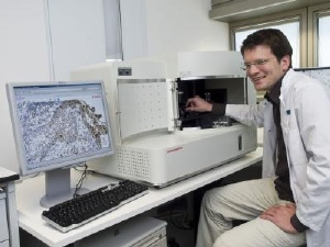
 For the first time a high-tech robot automatically reproduces and evaluates tissue slices only micromillimeters thick (right: Dr. Niels Grabe, research director at the TIGA center). Credit: Rothe
The Hamamatsu Tissue Imaging and Analysis (TIGA) Center is a cooperative effort between the Institutes of Pathology and of Medical Biometry and Informatics at the University of Heidelberg and the Japanese company Hamamatsu Photonics. In addition, it belongs to BIOQUANT, the research center for quantitative biology at the University of Heidelberg. At its core is the imaging robot "NanoZoomer" from Hamamatsu Photonics: the robot scans the tissue slices and displays them on the monitor for researchers at ultra high resolution and in various planes.
"Technically, this has brought the fully automatic evaluation of tissue changes and approaches for new therapy within our grasp," states Professor Dr. Peter Schirmacher, Director of the Institute for Pathology at Heidelberg University Hospital. This would represent a new milestone in pathology.
Detailed images help understand diseases
Which proteins are formed to a greater degree in cancer cells? How is tumor tissue changed during radiation treatment? Thanks to the NanoZoomer's high-resolution images and special evaluation programs, researchers in the future will be able to evaluate tissue and cell samples more quickly and accurately and gain important new insights for therapy tailored to the individual patient, for example for breast cancer.
In the future, the robot will be able to determine changes in cells and tissue fully automatically. "The NanoZoomer represents a quantum leap in tissue research," says Dr. Niels Grabe of the Institute for Medical Biometry and Informatics and research director at the TIGA Center.
Virtual Tissue is modeled from data
The medical IT specialists use the NanoZoomer to evaluate huge quantities of data from tissues for their research. For example, Dr. Niels Grabe and his team used data to model virtual skin tissue. "On a computer model of human skin tissue we can test whether certain substances are toxic, for example," explains Dr. Grabe. "In the future, this could make it easier to develop potential new drugs."
Hamamatsu recognized the many possible applications early on, so that new technological markets have now been opened up for them. "We are happy to have found two partners in the Heidelberg Institute of Pathology and the Institute of Medical Biometry and Informatics with whom we can develop concrete clinical uses and new applications for research," said Hideo Hiruma, Managing Director of Hamamatsu Photonics, Japan.