Nov 25 2008
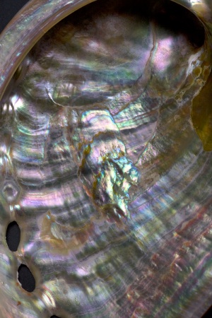
In addition to its iridescent beauty, mother of pearl, or nacre, the inner lining of the shells of abalone, mussels and certain other mollusks, is also renowned for an amazing strength and toughness that has been a long-standing mystery. Now, scientists have brought to light a new aspect of nacre's nanostructural architecture using the polarized x-ray beams and nanoscale imaging capabilities of the Advanced Light Source (ALS), a national synchrotron facility at the U.S. Department of Energy's Lawrence Berkeley National Laboratory (Berkeley Lab).
 Mother of pearl
Says Pupa Gilbert, a physics professor at the University of Wisconsin-Madison who led this research, "Nacre is a biomineral composed of thin layers of crystalline aragonite tablets separated by even thinner layers of organic material. Our studies have revealed that the aragonite tablet crystals in nacre are misoriented with respect to each other. This unique structural arrangement was a surprise and could play a role in nacre's remarkable resistance to fracture."
Approximately 95-percent of nacre's composition is aragonite, a hard but brittle calcium carbonate mineral, yet nacre is 3,000 times tougher than aragonite. No human-synthesized composite can outperform its component materials by such a wide margin. Hence nacre has been intensely studied, particularly in the ceramics and nanotechnology industries where brittleness remains a major limitation. The goal is to learn how nacre formation occurs in nature so that new microstructures might be engineered for ceramics and other materials that would increase their toughness. However, researchers in the past lacked the tools needed to study the dynamics of nacre formation at the nanoscale. Now, new capabilities at the ALS, including polarized beams of x-rays, have made this possible.
Gilbert and her colleagues reported their findings in the Journal of the American Chemical Society, in a paper entitled: "Gradual Ordering in Red Abalone Nacre." Co-authoring the paper with Gilbert were Rebecca Metzler, Dong Zhou and Susan Coppersmith, of the University of Wisconsin, and Andreas Scholl, Andrew Doran, Anthony Young, Martin Kunz and Nobumichi Tamura of Berkeley Lab.
Under the probing polarized light of PEEM-3, Gilbert and her colleagues obtained measurements of aragonite crystal orientation, tablet size and tablet stacking direction. These measurements revealed that different orientations of the aragonite crystals result in different tablet growth rates. In turn, these different table growth rates give rise to self-ordering, which progressively selects faster and faster growing tablets. Furthermore, this self-ordering takes place gradually over a distance of approximately 50 microns.
"Our results suggest that at least one aspect of nacre formation, the self-ordering of the aragonite tablets occurs independently of biological or organic-molecule control," said Gilbert. "Although this discovery shakes the generally accepted idea of organic-mineral templation in biomineralization, it is typical of evolving kinetic systems in nature to select for the fastest growing elements of a material."
From these results Gilbert and co-author Coppersmith hypothesized a "competition for space" theoretical model of nacre assembly, based on the gradual, kinetic selection of faster tablet growth rates and a small probability of nucleating new crystal orientations. The results of their model were found to be in remarkable agreement with side-view and in-plane experimental data.
One crucial piece that was still missing from the picture of nacre formation that Gilbert and her coauthors were putting together was information of the quantitative extent of the aragonite crystal misorientation both near and far from the boundary between nacre and the prismatic outer layer of the shell. According to Gilbert's hypothesis, the degree of orientational order of the aragonite tablets should increase with distance from the nacre-prismatic boundary. For this information, she turned to ALS Beamline 12.3.2, the x-ray microdiffraction facility.
Explains coauthor Tamura, who manages the x-ray microdiffraction beamline, "Our facility is capable of measuring with high spatial resolution and accuracy the orientation of individual aragonite tablets in nacre using a technique called white beam x-ray microdiffraction. For this study, we were able to do the measurements in a few hours and the crystal orientation numbers came out once we analyzed the data - the angular spread of the aragonite crystals misorientation goes from 24 degrees at the prismatic boundary down to 6 degrees a few tens of microns away."
The next step in this research, Gilbert says, is to extend the nacre study from red abalone, which is a gastropod mollusk, to the shells of other mollusks, including bivalves, such as certain oysters and mussels, and cephalopods, such as nautilus. Nacre in red abalone and nautilus is of a type known as "columnar," while the nacre in bivalves is of a type known as "sheet." It is already known that the formation mechanism for sheet nacre differs from that for columnar nacre.
Says Gilbert, "We are now working to acquire more PEEM-3 data on other nacre-forming shells, and simultaneously develop two separate theoretical models for nacre formation, one for sheet nacre and one for columnar nacre. These will be much more sophisticated models than what we have now."
This research was supported by awards from both the National Science Foundation and the U.S. Department of Energy's Office of Science.