Polymers are materials composed of repeated chains of molecules. Both natural and synthetic polymers possess many important properties that are influenced by the conformation and composition of monomers of individual macromolecules. One of the versatile analytical tools used for the nanoscale characterization of polymeric materials has been atomic force microscopy (AFM).

Image Credit: Egorov Artem/Shutterstock.com
AFM can help to understand the temporal shift in the morphology of polymers.
Polymer microstructures play an essential role in the functioning of flexible optoelectronic devices. The semiconducting organic polymers used in these devices are closely associated with transporting charge carriers and excitons, which determine the performance of these devices.
The relationship between carrier mobility and polymeric nanostructures has been previously reported using X-ray diffraction studies. Some of the tools used to characterize polymer thin films are scanning probe microscopy (SPM) and scanning tunneling microscopy (STM). Both these techniques have different approaches and operate under different conditions. These methods have issues related to the resolution of images obtained.
AFM is a microscope that provides images of the micro-nano-structures of the surface of a polymer, with a molecular-scale resolution. This is beneficial for polymer engineers to create new polymers with customized properties.
The nanotip probe, attached to a cantilever, made up of silicon, silicon nitride, gold, etc., is an important sensor for AFM.
The resolution of AFM depends on the structure and material of the AFM nanotip. A nanotip probe operates in two modes, i.e., contact or non-contact mode.
AFM provides three-dimensional images of polymers at molecular and submolecular levels. Preparation of samples for AFM study is relatively simple as it does not require any staining or metal coating. Additionally, this is a non-destructive method.
Characterization of Polymer Thin Films Using Atomic Force Microscopy
AFM has been used to acquire images of a single strand of synthetic polymers in several instances. Previous research has shown that incorporating torsional tapping-mode AFM imaging enables the imaging of single molecules.
AFM equipped with T-shaped ultra-sharp carbon-whisker tips can image semicrystalline domains of polymers, such as polyethylene, with a resolution of 0.37 nm in air.
Silicon nitride probes with ultra-high resolution are also used to acquire images at a molecular scale to understand the order of semiconducting polymer structure. This technique enables the imaging of polymer thin films with a roughness of a few nanometers.
One of the advancements of AFM is that it can operate at an increased temperature, which permits detecting in-situ polymer crystallization.
AFM is capable of observing crystal melting, growth, reorganizations, and assembly, which help understand the mechanism behind the growths of spherulites. It also helps evaluate crystallization parameters, such as temperature, sample thickness, and chemical composition, which are associated with crystallization kinetics.
In the study of polymer crystallization, the AFM probe tip must be contracted to analyze the crystal morphology throughout the scanning. This tool enables acquiring an image with sub-10 nm resolution.
AFM Coupled with Infrared Spectroscopy to Characterize Polymer Thin Films
Often AFM is coupled with other microscopies to study the composition of the thin polymer films. Recently, scientists have coupled AFM with tunable IR radiation (AFM-IR) to detect a polymer's photothermal effect. This method can also evaluate the chemical information down at a nanoscale resolution.
AFM-IR offers a controlled resolution of sub-diffraction by closely monitoring the diversion of an AFM nanotip that is in contact with the sample surface.
While scanning, the tip gets deflected by the thermal expansion of the sample after absorbing the infrared pulse.
The photothermal temperature of the sample is sensed by the AFM cantilever, and the cantilever deflections are measured using Fourier techniques. This technique can also evaluate the mechanical properties of the polymer samples.
Recently, researchers have proposed characterization of ultra-fine particles, which requires highly sensitive equipment, by AFM-IR associated with Lorentz contact resonance (LCR) imaging mode.
This advancement enables the characterization of ultra-thin films around 5 nm thick. AFM-IR is also applied via another mode called scanning thermal microscopy (SThM).
In this system, the special tip scans the surface of the polymer sample via contact mode. The change in the resistance is monitored by a Wheatstone bridge circuit and the output is generated as an image.

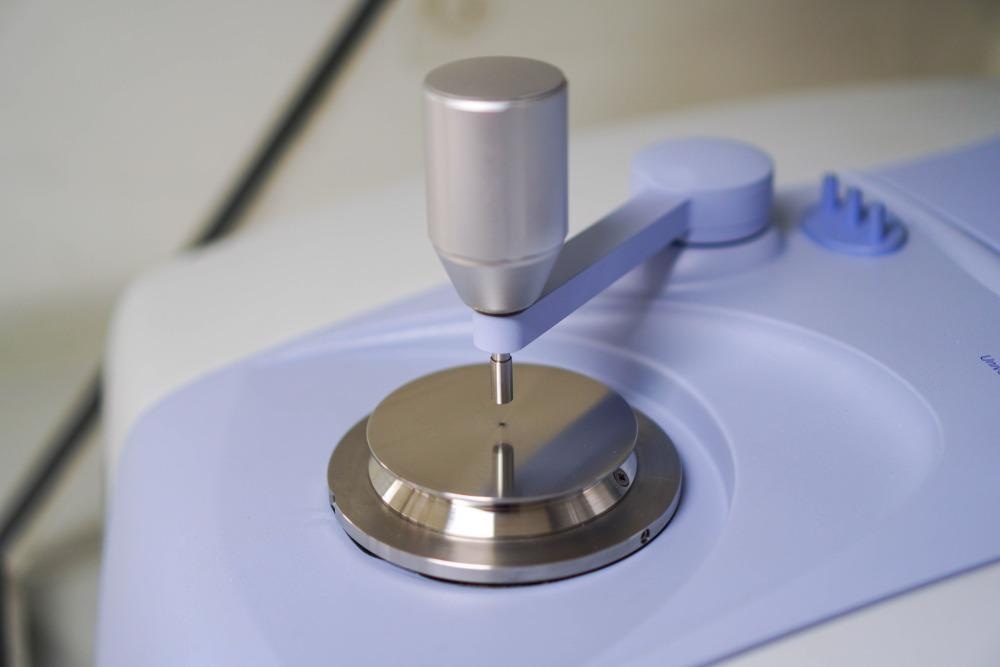
Image Credit: boonchok/Shutterstock.com
AFM Coupled with Confocal Raman Microscope to Characterize Polymer Thin Films
AFM coupled with a confocal Raman microscope (CRM) has been used to study the composition of polymeric film blends. This combination reveals the morphological characteristics of polymers at a nanoscale dimension.
It is also associated with analyzing the composition of various polymer thin film blends. Importantly, the topographic information and local stiffness of the polymeric film can be instantaneously obtained when samples are studied under AFM operated in Digital Pulsed Force Mode (DPFM).
The main advantage of this form of AFM is that it permits the material-sensitive classification of heterogeneous materials.
Scientists studied thin films of poly (methyl methacrylate) (PMMA) polymer, which is blended with two different styrene-butadiene rubbers using an AFM-CRM analytical tool.
PMMA is also known as engineered plastic and behaves as a glassy polymer at room temperature. Although AFM alone provided morphological characteristics of samples at a nanoscale, Raman spectroscopy elucidated the chemical composition of the polymeric film.
Tip-enhanced Raman scattering (TERS) utilizes local surface plasmon resonance effects at the probe tip to elucidate physicochemical information of a sample. This tool can characterize one-dimensional as well as two-dimensional polymer samples.
The confocal microscopy offered the spatial distribution of the molecules at various phases with a resolution of 200nm. The main advantage of using AFM-CRM is that it provides images of topographically different structures as well as the chemical composition of the polymeric samples.
Continue reading: Using AFM to Study Nanoemulsions.
References and Further Reading
Nguyen-Tri, P. et al (2020) Recent Applications of Advanced Atomic Force Microscopy in Polymer Science: A Review. Polymers.12(5), 1142. Available at: https://doi.org/10.3390/polym12051142
Korolkov, V.V. et al. (2019) Ultra-high resolution imaging of thin films and single strands of polythiophene using atomic force microscopy. Nature Communication. 10, 1537. Available at: https://doi.org/10.1038/s41467-019-09571-6
Schmidt, U. et al. (2005) Characterization of Thin Polymer Films on the Nanometer Scale with Confocal Raman AFM. Polymer Spectroscopy.230 (1). pp. 133-143. Available at: https://doi.org/10.1002/masy.200551152
Disclaimer: The views expressed here are those of the author expressed in their private capacity and do not necessarily represent the views of AZoM.com Limited T/A AZoNetwork the owner and operator of this website. This disclaimer forms part of the Terms and conditions of use of this website.