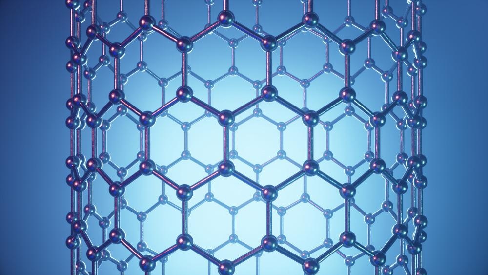
Atomic force microscopy (AFM) is a standard imaging technique for the structural characterization of surfaces in different fields of materials science, surface science, and biology. Carbon nanotubes have shown tremendous promise in the design and structure of AFM tips and probes.

Image Credit: Rost9/Shutterstock.com
The tip of an AFM probe is determined by the machine's lateral resolution, which in turn, determines its sensitivity.
The radius of curvature at the apex of conventional microfabrication methods is generally less than 10 nm. While developing smaller tips is a priority for AFM researchers, they also aim to develop tips that stay longer, accurately depict complicated surface topographies and are mechanically non-invasive.
Carbon Nanotubes and their Application
Carbon nanotubes (CNTs) have great potential in many research and industrial applications.
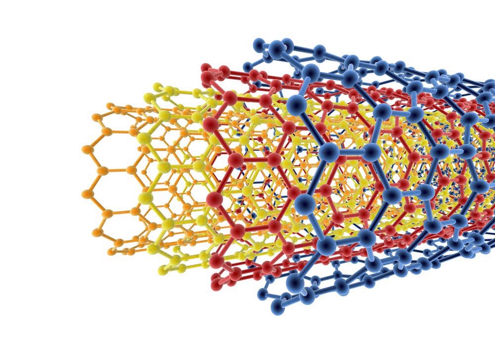
A single-wall carbon nanotube (SWCNTs) can be defined as a graphite sheet folded into a nanoscale tube or with additional graphene tubes around the core of an SWCNT, which are referred to as multi-walled CNTs (MWCNTs). The diameters of these CNTs range from fractions of nanometers to tens of nanometers, with lengths ranging from a few centimeters to several centimeters.
Chemical nerve agents can be detected by using thin-film transistors made from SWCNTs. They are reversible, detect concentrations as low as parts per billion (ppb), and are innately selective against hydrocarbon vapors and humidity interference signals. In a miniaturized capillary electrophoresis setup, a microdisk electrode made of MWCNTs and epoxy resin was utilized to measure the amperometric activity of bioactive thiols.
Carbon nanotubes are several times stronger than steel and other metals in terms of strength and are lighter in weight, making them a viable choice for structural reinforcement
Adding CNTs to cement-based materials has been proven in studies to boost performance considerably. When using CNTs in cement-based products, the ability of CNTs to be spread evenly is critical. CNTs are difficult to spread uniformly in cement-based materials because of their huge surface area, strong van der Waals force, and large aspect ratio. When a single MWCNT is isolated from its filling body, it is referred to as a CNT dispersion.
There are two sorts of approaches to dispersion techniques. The first is to use physical means, such as mechanical stirring, ultrasonic, and ball milling, to break down the material.
Carbon nanotube agglomerations can be broken down using ball milling, but the aspect ratio will be reduced, which negatively impacts the carbon nanotube's role in the matrix. Mechanical stirring includes magnetic and manual stirring, which is typically used in conjunction with ultrasonics to reduce the aspect ratio of carbon nanotube. The addition of functional carboxyl and hydroxyl groups and covalent or non-covalent bonds to the CNT's surface improves its wettability, resulting in the chemical dispersion of the CNT.
Benefits of CNTs in Atomic Force Microscopy
Carbon nanotubes have a nanoscale diameter and a high length/diameter ratio, unlike any other tubular structure. CNTs contain special properties such as a high aspect ratio which is used to image narrow and deep features.
Furthermore, they elastically buckle rather than break down when a high amount of force is applied to them, and for gentle imaging processes, they have low adhesion in tip-sample.
It is possible to employ controlled synthesis to manufacture every nanotube tip with the same structure and resolution because of the well-defined molecular architectures of nanotubes. As in previous structural approaches, a well-defined transfer function might be used to describe nanotube probes if the latter characteristic were achieved. SWNT bundle tips and multi-walled carbon nanotube (MWNT) tips for functionally sensitive imaging are used to get high lateral resolution images.
Nanotubes can have a diameter ranging from a few tens of nanometers to several hundreds of nanometers, depending on the number of concentric carbon shells used. There are several advantages to using carbon nanotubes as atomic force microscope probes, including the fact that they can have a one-nanometer diameter, strong mechanical qualities, and the ability to customize the tip end of the tube with chemical and biological probes.

Image Credit: Angel Soler Gollonet/Shutterstock.com
Carbon Nanotubes in AFM Systems
To physically contact and evaluate a sample surface, atomic force microscopy (AFM) uses an ultra-sharp tip. Using new nanotechnology processes, the technology for making AFM probe tips is rapidly evolving. A new generation of AFM probes is now easily accessible, with increased performance, higher quality, and novel materials.
Probes made of carbon nanotubes offer a new AFM imaging universe, boosting the probe's resolution and endurance, reducing the probe–sample forces, and broadening the AFM application areas.
Nanotechnology, surface engineering, and biotechnology would all benefit from this breakthrough. Using carbon nanotube tips in AFM has various benefits, such as performing gentle imaging and using low tip-sample adhesion. To bend elastically upon severe pressure load rather than breaking. Such properties make use of CNT in AFM systems very much appealing and beneficial.
The CNT-AFM probes were found to be very easy to use for regular imaging in Peak Force Tapping (PFT) mode. Additionally, it has been found that chemical vapor deposition (CVD) nanotube tips offer superior imaging capabilities for biomolecules than commercial tips or manually assembled nanotube tips when used in the same environment.
Challenges Faced During Usage of CNTS in AFM
When using a CNT-AFM probe for AFM imaging, there are a lot of crucial elements that must be regulated efficiently, but possibly the most significant is the applied force. An unstable contact is possible if too much force is exerted on the nanotube.
When it comes to Probe production, the length of the CNT and the orientation are crucial considerations, making these probes particularly challenging. Because of the strong lateral stresses, CNT-AFM probes are not suitable for imaging in contact mode for the reasons stated above. As a means of reducing forces (lateral), CNT-AFM probes are often used in tapping (mode).
Continue reading: FluidFM - Where Nanofluidics and AFM Meet
References and Further Reading
Endo, M., Hayashi, T., Kim, Y. and Muramatsu, H., (2006). Development and Application of Carbon Nanotubes. Japanese Journal of Applied Physics, 45(6A), pp.4883-4892. https://doi.org/10.1143/JJAP.45.4883
Slattery, A., Shearer, C., Shapter, J., Blanch, A., Quinton, J. and Gibson, C., (2018). Improved Application of Carbon Nanotube Atomic Force Microscopy Probes Using PeakForce Tapping Mode. Nanomaterials, 8(10), p.807. https://www.mdpi.com/2079-4991/8/10/807
Chin Li Cheung, J. H. (2000). Carbon nanotube atomic force microscopy tips: Direct growth by chemical vapor deposition and application to high-resolution imaging. PNAS. https://www.pnas.org/doi/full/10.1073/pnas.050498597
Wilson, N. and Macpherson, J., (2009) Carbon nanotube tips for atomic force microscopy. Nature Nanotechnology, 4(8), pp.483-491. https://doi.org/10.1038/nnano.2009.154
Popov, V., 2004. Carbon nanotubes: properties and application. Materials Science and Engineering: R: Reports, 43(3), pp.61-102. https://doi.org/10.1016/j.mser.2003.10.001
Xu, Z., Fang, F. and Dong, S., 2011. Carbon Nanotube AFM Probe Technology. Electronic Properties of Carbon Nanotubes,. https://www.intechopen.com/chapters/16843
Disclaimer: The views expressed here are those of the author expressed in their private capacity and do not necessarily represent the views of AZoM.com Limited T/A AZoNetwork the owner and operator of this website. This disclaimer forms part of the Terms and conditions of use of this website.