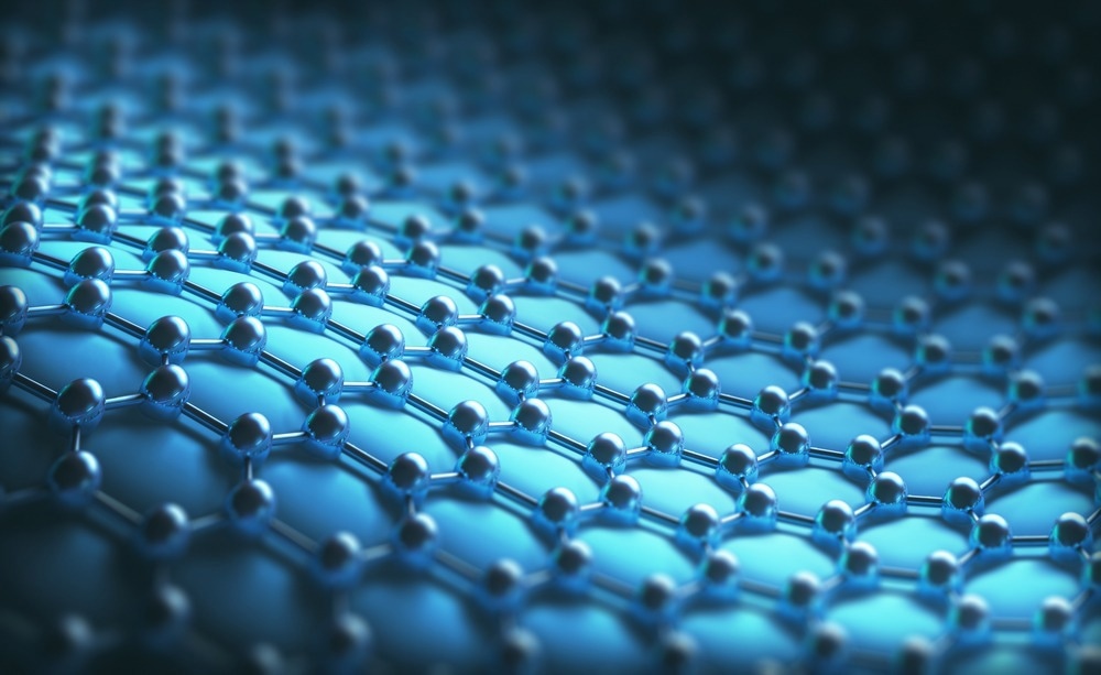
Engineering material surface properties can influence some of the most important characteristics of materials, such as corrosion resistance, adhesion, color, conductivity, biocompatibility, and many others.

Image Credit: ktsdesign/Shutterstock.com
The growing demand for high-performance materials boosts the significance of a range of surface chemical analysis techniques and their importance for developing new consumer products and devices.
Depending on the material composition, a surface layer typically consists of three to five atomic layers (1-2 nm thick). Thicker layers, with a thickness of up to 10 nm and one micron, are considered ultra-thin and thin films, respectively.
Why Are Surfaces Important?
The material's surface is the interface where the material interacts with the environment and other materials. Many issues, such as corrosion, adhesion, catalytic activity, wettability, friction, and others, associated with using modern materials in various industrial applications can be solved only by better understanding the physical and chemical interactions occurring at the material's surface or interface.
By understanding the materials' surface chemistry, scientists and engineers can alter or enhance the materials' characteristics through precisely controlled engineering and modification of surface properties.
Furthermore, surface chemical characterization is of great importance for a wide range of manufacturing processes, like semiconductor manufacturing, where even a minimal amount of surface contaminants can dramatically degrade the product quality and prevent it from meeting the desired quality standards. Thus, surface chemical analysis can be used for developing novel materials with advanced functionality and stringent quality control in materials processing.
Surface chemical analysis of materials is vital for understanding how materials interact at interfaces. Several surface chemical analysis techniques have greatly contributed to the advancement of surface science in the last several decades by enabling researchers to investigate the elemental composition and chemical bonds specifically on the surface of a wide range of materials.
Over the years, surface analysis methods have advanced with the needs of academia and industry as essential tools for R&D and quality assurance in many industrial and scientific research fields.
Principles of Surface Chemical Analysis
Surface chemical analysis employs analytical techniques where beams of electrons, ions, neutral atoms or molecules, or photons (ultraviolet, visible, or X-rays) are directed at the specimen surface and scattered or emitted electrons, ions, or photons are collected and analyzed. A complementary approach is to employ scanning-probe-based microscopic techniques where functional probes are scanned near the material's surface and surface-related signals are detected. Combining surface characterization techniques with ion milling permits scientists to examine subsurface properties and acquire 3D chemical composition depth profiles.
Electron Spectroscopy-Based Analytical Methods
Auger electron spectroscopy (AES) employs a focused, high-energy electron beam scanned over the ample surface to excite electron shell-to-shell transitions in the atoms of the surface layer. The excited atoms relax by emitting either X-rays or outer shell electrons (called Auger electrons). The kinetic energy spectrum of the Auger electrons is then analyzed and provides information about the elemental composition of the specimen.
The shallow escape depth of the Auger electrons ensures that the technique is specific only to the top few atomic layers of the sample material. Scanning Auger microscopy permits surface chemical mapping with lateral resolutions better than 1 μm. AES is highly suitable for analyzing metals and semiconductors and is widely used to characterize metal evaporation and deposition, corrosion, and welding.
X-ray photoelectron spectroscopy (XPS, also known as electron spectroscopy for chemical analysis or ESCA) employs soft X-ray radiation (with photon energies in the range of 200-2000 eV) to ionize electrons from the surface of a solid sample. The closely-related UV photoelectron spectroscopy technique employs vacuum UV radiation (with photon energies in the range of 10-45 eV) to excite the valence electrons in the analyzed material.
In both cases, the energy spectrum of the emitted electrons is characteristic of the binding energy of the material elements and the associated chemical bonds in the top few atomic layers of the material.
XPS and UPS are quantitive spectroscopic techniques that can provide information about the elemental composition, atomic concentrations, and chemical states of the elements present at the sample surface (analysis depth of approximately 3-5nm). The techniques can detect all elements except hydrogen and helium. The area of analysis is typically several tens of square millimeters.

Image Credit: Swill Klitch/Shutterstock.com
Ion Beams Probe Surface Chemistry
Secondary Ion Mass Spectrometry (SIMS) employs a focused beam of charged primary ions (typically gallium ions) to sputter atoms, ions, and molecules from the outermost atomic layer of the sample surface. The ion beam dose is adjusted to achieve minimal surface damage; hence the technique is considered non-destructive (known as static SIMS), although it still removes material from the surface. In dynamic SIMS, a higher ion dose is used to sequentially remove the surface layers, thus enabling chemical depth profiling.
The sputtered secondary ions are analyzed depending on their mass-to-charge ratio by a quadrupole mass spectrometer. Besides the secondary ions, the sputtering process generates large numbers of electrons, which can be used to produce an image of the sample surface similar to scanning electron microscopy.
SIMS spectra can also be acquired while scanning the primary ion beam across the sample surface, thus generating a high-resolution chemical composition map of the specimen. Importantly, the technique can detect all elements, including isotopes of the same element.
The combination of SIMS and XPS is a powerful analytical tool for comprehensive surface elemental mapping chemical state quantification. These techniques are widely used in microelectronics and semiconductor industries, chemical engineering, catalysis, and soft matter research.
Vibrational Spectroscopy for Surface Analysis
Vibrational spectroscopy enables the most accurate identification of surface species resulting from chemical reactions or molecular adsorption.
Fourier transform infrared (FTIR) and Raman spectroscopy are non-destructive techniques sensitive to changes in the molecular structure of the materials and widely used for simple and rapid investigation of molecular bonding in materials.
Both techniques rely on incident photon (typically ultraviolet to infrared) scattering and energy loss through exciting vibrational modes of the molecular bonds on the sample surface. The spectrum of the scattered photons is analyzed to identify the molecular species on the sample surface to make a spectrum.
Raman spectroscopy typically has a depth of analysis of about one micron, much larger compared to some of the other techniques described here. However, the technique can be combined with microscopic imaging methods to provide spatial information for the distribution of molecular species with micron-scale resolution. The vibrational spectroscopic techniques are especially useful when investigating the chemical properties of polymers and nanomaterials, such as graphene and carbon nanotubes.
References and Further Reading
Sykes, D. (2017) Surface Chemical Analysis. In: Kasap, S., Capper, P. (eds) Springer Handbook of Electronic and Photonic Materials. Springer Handbooks. Springer, Cham. Available at: https://doi.org/10.1007/978-3-319-48933-9_18
Erdoğan, G., et al. (2017) Surface Characterization Techniques. In Surface Treatments for Biological, Chemical, and Physical Applications (eds M. Gürsoy and M. Karaman). Available at: https://doi.org/10.1002/9783527698813.ch3
Baer, D. R., et al. (2010) Application of surface chemical analysis tools for characterization of nanoparticles. Anal. Bioanal. Chem. 396(3), 983-1002. Available at: https://doi.org/10.1007/s00216-009-3360-1
Disclaimer: The views expressed here are those of the author expressed in their private capacity and do not necessarily represent the views of AZoM.com Limited T/A AZoNetwork the owner and operator of this website. This disclaimer forms part of the Terms and conditions of use of this website.