May 27 2009
Rice physicist Huey Huang is on a quest to understand death -- or at least a little piece of it. Huang has spent the past 15 years studying the properties of cell membranes in an effort to unravel a mystery about cell suicide, a mystery that starts with a tiny hole.
 Rice physicist Huey Huang pioneered the use of bromine atoms (red) as markers in membrane studies. Last year, the technique helped Huang confirm the toroidal shape of pores that trigger cell suicide.
The hole is important because it's a trigger. It kicks off a process known as apoptosis, or cell suicide, and scientists want to understand apoptosis because of the role it plays -- or fails to play -- in cancer. In healthy bodies, defective cells are marked for an orderly death by apoptosis. These cells commit suicide and even have the courtesy to package their remains for convenient recycling. Why this happens is a mystery. How cancer cells avoid apoptosis is perhaps a bigger mystery, and one reason scientists want to crack the code on apoptosis is to find better ways to fight cancer.
To illustrate just how little scientists know about apoptosis, Huang, Rice's Sam and Helen Worden Chair of Physics and Astronomy, pulled down a leading cell biochemistry textbook from his office shelf and opened to the chapter on apoptosis.
"This is all," he said, leafing through a handful of pages that make up the entire chapter. "We really understand very little about it."
Thanks to a breakthrough in Huang's lab late last year, scientists now understand a key feature of apoptosis: They know the shape of the hole, or pore, that triggers it.
Huang's groundbreaking work recently earned him a four-year grant from the National Institutes of Health. With the grant, Huang will mark two decades of continuous NIH funding for pioneering research in biophysics.
Apoptosis is triggered when a hole is punched through a membrane that walls off the mitochondria inside a cell. The mitochondria are the cell's internal power centers, the place where the cell produces the energy necessary to live.
In cells marked for suicide, a protein called Bax gets a signal -- nobody knows what the signal is -- and starts punching holes in the outer wall of the mitochondria. Molecules leak out of these pores, bind with proteins and kick off a process that ends with "executioner" proteins systematically dismantling the entire cell.
"It's the very first stage," Huang said of Bax's pore formation. But knowing that Bax forms pores and understanding how it forms pores are two different things. For one thing, scientists have only known that proteins could form pores in membranes since 1996, and most of the research on the structure of these pores has been done on proteins that are different from Bax. These other pore-forming proteins can fold themselves into barrel-like cylinders, and scientists have decoded their pore structures. But Bax can't form pores this way because it cannot wrap into a barrel.
The sack-like membranes that surround cells and mitochondria are thinner than a garbage bag. They're made of two sheets of fatty acids that stick together very tightly, like pieces of cellophane tape. The fatty acids in the membrane help form a watertight seal for the contents inside.
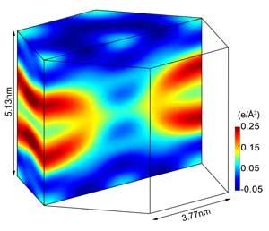
In 1996, Huang and his graduate students proposed a new idea about the way proteins might form pores in membranes. They suggested that certain proteins, including Bax, react with the bilayered membrane in such a way as to cause it to curve, forming a rounded hole like the one in a doughnut. Late last year, Huang, students and longtime colleague Lin Yang of Brookhaven National Laboratory in Upton, N.Y., used the National Synchrotron Light Source at Brookhaven to take hundreds of painstaking X-ray diffraction images of pores formed by pieces of Bax. They confirmed the toroidal, or doughnut-shaped, hole, settling the debate about how Bax forms holes in membranes.
Huang said the group is now turning its attention to a more difficult investigation. The group is trying to work with the entire Bax protein to find out what causes it to start making holes in the first place.
"Compared to our previous work on peptides (pieces of Bax), working with the whole protein is much harder," Huang said.
But Huang's group is well-positioned for the new task. It is the only research group in the world that's focused solely on studying "protein-induced structure in lipid bilayers." And as such, the group has pioneered many of its own techniques for investigation. One ingenious example of this is the group's bromine marker technique. Bromine atoms give off a unique diffraction signal when they're exposed to X-rays, and Huang, Yang and colleagues helped infer the shape of the toroidal pore last year based on the way bromine atoms were distributed through their samples.
"It made for some really beautiful images," Huang said. "I've often seen our 1996 images used in talks at conferences, but these are much more compelling. I suspect these new images will be used in many a talk in the coming years."