Chromosomal dissection enables separation of DNA from cytogenetically identified region to produce genetic probes for exhibiting fluorescence in situ hybridization, a method that is popular in cyto and molecular genetics study and diagnostics.
 AFM
AFM
Several reports are available that provide information regarding methods of microdissection such as laser beam or glass needle to acquire specific probes from metaphase chromosomes.
The traditional dissection methods tend to have many disadvantages as it requires numerous chromosomes for manufacturing a probe. Also, due to the large size of microneedles, the traditional methods are not apt for single chromosome analysis. Hence it is important to have new dissection techniques that can provide advancement in chromosomal research at the nano level.
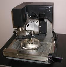
The atomic force microscope (AFM) is used as a tool for single chromosomes nanomanipulation to produce single genetic probes that target particular cells. This method integrates nanomanipulation and molecular techniques to enable amplification and nanodissection of chromatidial and chromosomal DNA.
A cross-sectional examination of dissected chromosomes displays 20 nm and 40 nm deep incisions. Degenerate oligonucleotide-primed polymerase chain reaction (DOP-PCR) is applied to amplify and label stand-alone, individual chromosomal portions and later to hybridize to interphasic nuclei and chromosomes.
Atomic force microscope can be used to view and control biological substances with high precision and resolution. The fluorescence in situ hybridization (FISH) carried out with DOP-PCR devices as test probes have been effectively experimented in interphasic nuclei and avian microchromosomes.
Chromosome nanolithography has a resolution higher than that of light microscopy. It can be helpful for constructing chromosome band libraries and mapping of the molecular cytogenetic mapping used for study of genetic diseases.