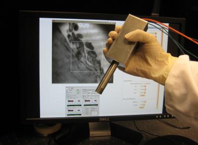
A team of scientists from the University of Texas at Austin has devised a handheld microscope that shows promise in decreasing the time consumption to detect oral cancer.
 A team of American researchers have created a portable, miniature microscope in the hope of reducing the time taken to diagnose oral cancer. (Credit: Dr. John X.J. Zhang, Department of Biomedical Engineering, the University of Texas at Austin.)
The microscope with a 20-cm-long and 1-cm-wide tip holds potential to be utilized as a surgical guidance instrument or as a real-time oral cancer diagnostic tool. This probe can also be used by dentists to examine premature cancer cells.
The novel tool generates full 3D images of areas within a tissue surface by illuminating the sample areas with a laser. It can deliver a large field-of-view by taking a sequence of images and layering them on top of one another.
The device’s major component is a micromirror, which is used in fiber optics and barcode scanners. A microelectromechanical system is used to control the micromirror, which in turn enables the laser beam to monitor a surface in a programmed manner. These micromirrors are affordable, can be produced easily, and can be integrated easily into electronic systems for a variety of imaging operations. These features make them a crucial part of the microscope.
The research team at the University of Texas at Austin together with its commercialization partner, NanoLite Systems, intends to conduct clinical studies to get the FDA approval. Through fine tuning, they believe that the cost of the device can be reduced to one-fourth the price of the present microscopes utilized in diagnosis.
The innovative microscope has been reported in Journal of Micromechanics and Microengineering.