Oct 29 2012
Using something called “inertial microfluidics,” University of Cincinnati researchers are able to continuously and selectively collect rare cells, such as circulating tumor cells, based on their size vs. other biomarkers. This could reduce analysis time and increase selectivity while reducing reliance on antibody-based testing in clinical tests.
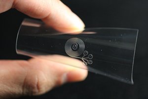
 Another view of microfluidic device with spiral microchannels and four outlet ports which separates blood into different streams.
UC ingot At the Sixteenth International Conference on Miniaturized Systems for Chemistry and Life Sciences (microTAS) to be held Oct. 28-Nov. 1, in Okinawa, Japan, University of Cincinnati researchers will present four papers, including one detailing improvements in rare cell isolation and one detailing improvements, in terms of cost and time, of common blood tests.
Ian Papautsky, associate professor in UC’s School of Electronic and Computing Systems (SECS), part of the College of Engineering and Applied Science, and a UC team are leading these research efforts.
In a paper titled “Continuous Rare Cell Extraction Using Self-Releasing Vortex in an Inertial Microfluidic Device” by Papautsky and co-authors Xiao Wang, UC doctoral student, and Jian Zhou, research associate, a new concept for separation of rare cells, such as prostate cancer cells or circulating tumor cells, using microfluidics, is detailed.
“Last year we showed we can selectively isolate prostate cancer cells, but only by running small sample volumes one at a time. Now we show that we can do this continuously,” Papautsky said. “This is exciting because it allows for an entire blood draw to be processed, in continuous matter, in a shorter period of time.”
These blood draws can be used to identify tumor cells for diagnostic or prognostic purposes. “Our approach is based purely on size. It doesn't rely on antibodies, which is important because not all cancer cells express antigens. So, if the cancer cells are, let's say, larger than 20 microns, we'll extract them,” he explained.
Size-dependence of particle trapping
The most common approach for looking for these circulating tumor cells is via a system that uses a selection using antibodies to detect antigens. “We could also use our device to prepare samples for systems that use antibody-based selection.” This combined approach could potentially help reduce occurrence of false positives while significantly increasing the accuracy of the antibody-based tests.
Another area in which this device could be useful is in working with cell cultures. “If you have a mixture of multiple cells where some cells are small and other cells are big, we could separate these cell populations very easily,” Papautsky explained. “Anytime you need to separate based on size, we can do it using inertial microfluidics.”
The advantage of inertial microfluidics in cell separation is that it can be done easily and without cumbersome equipment. This research is leading to an entirely new generation of testing capabilities which particularly lend themselves to direct use in the field and in physicians’ offices in just about any country and any economic setting.
In another paper, titled “Sorting of Blood in Spiral Microchannels” Papautsky and doctoral student Nivedita Nivedita demonstrate continuous sorting of blood utilizing inertial microfluidics via a simple passive microfluidic device. Papautsky’s lab has been developing the concept of using inertia to manipulate cells and particles during the last few years. “It's truly different and innovative because these microfluidic devices are really low cost while offering very high throughput,” said Papautsky.
The device is, essentially, a clear, plastic, flexible square that is relatively small in size, at about a half an inch across, but big in concept. “With this particular device we can take a drop of blood, put it in the input port in the center, and separate,” Papautsky explained. The device contains four outlet ports which separate the blood into different streams, allowing the collection of outputs containing dilute plasma, red blood cells and white blood cells.
spiral microchannels
“There are a lot of clinical diagnostic tests that are based on blood,” he said. One of the most common tests that are done in a hospital is the complete blood count (CBC). Through this test, a wide range of conditions like anemia, malaria or leukemia are diagnosed. “In all of these diagnostic tests, blood must be separated into its components, and that's what this device does,” Papautsky explained. “So, instead of using a big centrifuge to do it, we can do it with this little device.” Using the microfluidic device allows for a diagnosis in less time in a much easier fashion.
This quick, low-cost way of running a diagnostic test could potentially be used in a resource-limited setting. “One of the issues that I hear from my colleagues who work in these areas that do tests is that they have equipment,” he said, “but don't always have personnel or stable power to operate them. So in places like India, Africa or Central America, our devices could be useful.”
This work was supported by the DARPA Micro/Nano Fluidics Fundamentals Focus (MF3) Center at the University of California at Irvine.