Jan 8 2013
How do viruses attach to cells? How do proteins interact and mediate infection? How do molecular machines organize themselves in healthy cells? How do they differ in diseased cells? These are the types of questions National Institutes of Health researchers ask in the recently established Living Lab for Structural Biology, questions they strive to answer through the most sophisticated of imaging techniques.
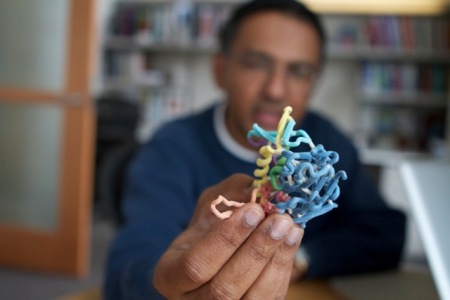
 Sriram Subramaniam, Ph.D., holds a 3D model of gp120, part of a protein complex displayed on the surface of HIV. The Living Lab aims to develop automated workflows to rapidly determine the structures of small dynamic protein complexes of this kind.
The Living Lab is an innovative partnership between NIH and FEI, an Oregon-based instrumentation company that manufactures advanced microscopes. FEI brings to the table invaluable assistance in developing and customizing electron microscopes for biological applications. Using that cutting edge technology, scientists in the Living Lab, unencumbered by any pressure to patent or otherwise protect discoveries for commercial purposes, can proceed purely driven by scientific and biomedical puzzles. Success of the Living Lab, which is on the NIH campus in Bethesda, Md., will rest on that collaboration between the government and the private sector—and the idea that answering scientific questions and technical advancement go hand in hand.
“We want to navigate our way into cells and into viruses,” said Sriram Subramaniam, Ph. D., director of the NIH component of the Living Lab. “We would like to be able to describe the function of complex things, such as whole cells or infectious viruses, in terms of their molecular make-up, and try to figure out how they work.”
The Living Lab’s advanced imaging technology allows researchers to tackle previously unanswered questions in structural biology by creating three-dimensional shapes of various molecular machines. Visualizing tiny details is a step toward understanding the molecular origins of disease. “The prospects for studying structures of a broad spectrum of medically relevant complexes at minute resolutions has changed dramatically in recent years with advances in structural biology,” said Subramaniam. “Our goal with the Living Lab is to capture the synergy between all of these methods including the latest advances in cryo-electon microscopy to extend these advances to key scientific challenges in modern structural biology.”
Subramaniam, who earned his doctorate at Stanford University and did post-doctoral work at the Massachusetts Institute of Technology in chemistry and biology, directs the research activities of the Living Lab, in close consultation with other team members from FEI and from the National Institute of Diabetes and Digestive and Kidney Diseases.
The Living Lab is, in many ways, an evolution of work he has long led in the Laboratory of Cell Biology in NCI’s Center for Cancer Research. Subramaniam and his colleagues are at the forefront of their field, using high-powered electron microscopes to create 3-D maps of proteins, viruses, and cells, including HIV and cancer cells.
What the Living Lab offers is more capacity, more expertise, and more technology. It is, says Subramaniam, an opportunity for the NIH to make real breakthroughs, imaging proteins at higher resolutions, and understanding the structures of more challenging molecules. “This lab is unique because the science is so interdisciplinary. The things that we do, the science here, benefits from expertise drawn across the campus and across the world.”
A Titan Krios transmission electron microscope, one of the world’s most powerful commercially-available electron microscopes, is at the heart of the Living Lab. The microscope, a two-story, two-toned box that holds and insulates temperature-controlled components, rises from floor to ceiling. The Krios can collect data with a high degree of automation and can be operated remotely, taking pictures of proteins and other biological assemblies on its own for days at a time, without human intervention.
Cryo-electron microscopy of this type has gained popularity in structural biology research because it allows for the observation of specimens that have not been stained or fixed in any way, presenting them in their native environment. Previous technologies, Subramaniam explains, have been analogous to “destroying a house in order to describe the building that used to be there.” With this enhanced technology and better software, scientists can capture important details before proteins are damaged, allowing much more detail to be resolved.
Subramaniam describes his team as basic scientists looking to figure out how proteins work. But the Living Lab is pushing that forward, broadening the scope of the types of protein and protein assemblies that can be studied. As they push the capabilities of the technology, the team will be moving ever closer to answering those big, biological questions, one protein at a time.