May 26 2014
Biologists and doctors rely heavily on incubators and microscopes. Now the Fraunhofer Institute for Biomedical Engineering IBMT has come up with a novel solution that combines the functions of both these tools in a compact and extremely small-scale system.
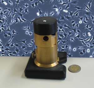
It is ideally suited for time-lapse examination over a number of weeks and for automatic observation of cell cultures. The incubator microscope is no bigger than a soda can and costs 30 times less than buying an incubator and a microscope separately. It will be on display for the first time at MEDTEC in Stuttgart (Hall 7, Booth B10).
 No bigger than a soda can, the small-scale incubator microscope is a space-saving and cost effective solution for time-lapse observation of cell cultures. (© Fraunhofer IBMT)
Cells play a prominent role in biology and in medicine. Just like humans, they need nutrients to survive. Cultivating human and animal cells requires parameters such as temperature and humidity to be specified with absolute precision and maintained at an even level over long periods of time. Time-lapse observation over a period of some weeks can be particularly valuable, since a lot happens in that time in terms of cell reproduction and differentiation. Until now, the usual technique to make these sorts of observations has been to use small incubators in combination with conventional microscopes. This takes up about one square meter of space, making operating several such systems alongside each other an inefficient process. There is a need for innovative solutions that will significantly reduce the space needed and the costs involved – without compromising the quality of the cultivation and of the microscope images recorded.
At MEDTEC researchers from the Fraunhofer Institute for Biomedical Engineering IBMT in St. Ingbert will be showcasing their small-scale incubator microscope solution. This system can be used for time lapse observation of cell cultures as well as to gather fluorescent images at different wavelengths. It includes a small-scale incubation chamber and control electronics to ensure defined cell culture parameters. Cells grow on the floor of the miniaturized incubation chamber on a thin, replaceable glass plate and are supplied with a constant stream of nutrients. The only parameters that need to be kept constant within the incubator are the temperature and the nutrient supply flow rate. All in all, the small-scale incubator microscope is extremely good value and allows for many units to be operated in parallel in a very compact space. And despite its space-saving design, the system yields images that are almost as good as those of the big microscopes.
Prototype versions are already in use in a variety of research projects. “The system is stable and can be used for time-lapse observation spanning several weeks,” says Dr. Thomas Velten, head of the Biomedical Microsystems department. The device continuously gathers data and saves them to a computer. Images can be accessed at any time and analyzed using the appropriate image processing software.
“Our customers get a biomedical analysis tool of the highest quality – well priced, space-saving and tailored to their needs,” says Velten. The incubator microscope is suited to a wide variety of applications, for instance examining the reaction of cells to nanoparticles or toxic agents in the environment. Another current application is stem cell research. “The system is compact, mobile, extremely efficient and fully automatic in operation,” concludes Velten.