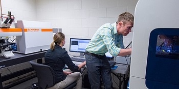
The Biomechanics and Biomimetics Research Laboratory at the University of Colorado Boulder uses a Renishaw inVia™ confocal Raman microscope to characterise biological tissues and biomaterials.
 Users from the Virginia Ferguson Laboratory at the University of Colorado Boulder work with the Renishaw inVia Raman system interfaced to the Hysitron TriboIndenter system
The system has been interfaced with a nanoindenter system from Hysitron Inc, Minneapolis, USA, to provide combined chemical and mechanical information on samples. Professor Virginia (Ginger) Ferguson is an Associate Professor of Mechanical Engineering at the University of Colorado who focuses on the mechanical behaviour, and underlying materials science, of biological tissues and biomaterials. Her group investigates the local mechanical properties of materials that are biological, biomimetic or used in biological tissue replacement (e.g. for tissue repair and regeneration), correlating these properties with chemical and structural information obtained from Raman and other techniques.
More specifically, Professor Ferguson’s research emphasises: the experimental study and finite element modelling of native osteochondral (bone-cartilage) tissues and of materials for osteochondral tissue regeneration; bone tissue properties in mouse models that are affected by unloading and microgravity, aging, and/or metabolic disease (e.g. kidney disease); and tissues involved in successful pregnancy outcomes in humans (e.g., the cervix and chorioamnion—the ‘amniotic sac’).
The group has developed protocols and analytical solutions to collect and evaluate nanomechanical property measurements of bone, calcified cartilage and soft tissues (e.g. cartilage). They also evaluate collagen fibril orientation and alignment using secondary harmonic generation imaging and evaluate mineral volume fraction in mineralized tissues using quantitative backscattered electron imaging. The team collaborates with other groups to test materials for a wide range of engineering applications including materials for batteries/energy storage, microelectro-mechanical systems (MEMS) devices, advanced polymers and other complex material systems which makes it a varied and interesting laboratory to work in.
Pairing Renishaw’s inVia Raman microscope with the nanoindenter from Hysitron results in a combined instrument that provides a greatly enhanced characterisation capability, complementing existing spectroscopies. Professor Ferguson said, “This unique system provides the capability to investigate mechanical properties and mechanical behaviours in parallel with imaging the chemical constituents within the materials being tested at exactly matched sites / locations with ~1 µm resolution. We use the information on a material’s chemistry to elucidate changes in mechanical properties or to explain mechanical properties within single sites or when mapping over 2D regions. Moreover, mapping reveals gradients or heterogeneity of material chemistry and crystal structure. We can also uniquely evaluate chemical changes that occur following pressure induced phase transformations (that could be applied by nanoindentation, for example).”
The inVia offers a degree of customization that is not available on other systems. More importantly, we like the manner in which the inVia collects Raman spectra compared to other systems. We are the first to have joined the Hysitron nanoindenter with any Raman spectroscopy tool and have had technical challenges to overcome. I have been exceptionally pleased with the level of customer support we have received from Renishaw during the development of our unique combined instrument. Renishaw has been fantastic to work with.
Professor Ferguson
Further information on the work of the group may be found at Professor Ferguson’s web site, the Biomechanics and Biomimetics Laboratory. For further details of Renishaw’s inVia confocal Raman microscope, please visit www.renishaw.com/invia