Apr 24 2019
A complex cable system of muscles and tendons hold the human body together. This system is designed by nature to be highly stretchable and strong.
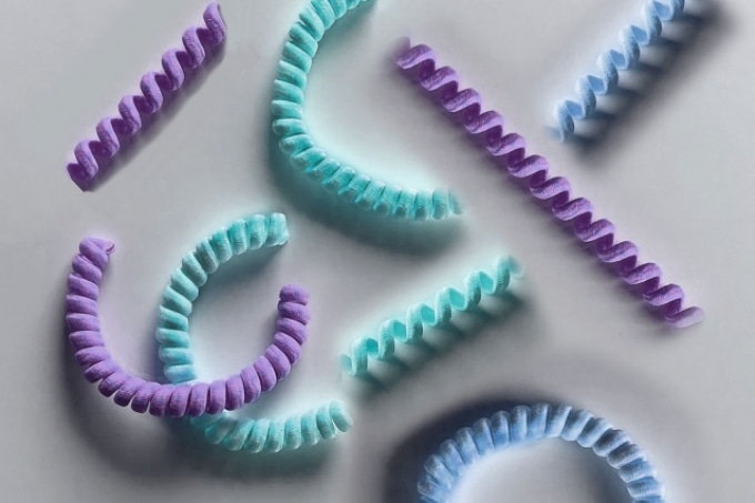
 MIT engineers have designed coiled “nanoyarn,” shown as an artist’s interpretation here. The twisted fibers are lined with living cells and may be used to repair injured muscles and tendons while maintaining their flexibility. (Image credit: Felice Frankel)
However, if an injury is caused to any of these tissues, especially in a crucial joint like the knee or shoulder, then weeks of limited mobility and surgical repairs will be required to enable them to heal completely.
Now, a team of engineers at MIT has developed a new kind of tissue engineering design that could allow a flexible range of motion in damaged muscles and tendons at the time of the healing process.
The researchers have designed tiny coils that are lined with living cells, which according to them can serve as flexible scaffolds for repairing injured tendons and muscles. Fabricated from a countless number of biocompatible nanofibers, the coils are firmly twisted into coils looking like tiny nautical yarn, or rope.
The yarn was lined with living cells, including muscle cells and mesenchymal stem cells, which grow naturally and easily align beside the yarn, into designs akin to muscle tissue. The scientists discovered that the coiled configuration of the yarn helps in keeping the cells alive and thriving, even as the yarn is bent and expanded many times.
In the coming days, the engineers believe that physicians can possibly line the injured muscles and tendons of patients using this novel flexible material, which would be lined with the same kind of cells making up the damaged tissue. A patient’s range of motion could be maintained by the stretchiness of the yarn, while fresh cells continue to grow to substitute the damaged tissue.
When you repair muscle or tendon, you really have to fix their movement for a period of time, by wearing a boot, for example. With this nanofiber yarn, the hope is, you won’t have to wearing anything like that.
Ming Guo, Assistant Professor, Department of Mechanical Engineering, MIT.
Guo and his coworkers have recently reported the results of their study in the Proceedings of the National Academy of Sciences. His co-authors at MIT are Yiwei Li, Satish Gupta, Yukun Hao, and Jiliang Hu. The group also includes Yaqiong Wang, Fengyun Guo, Yong Zhao, and Nü Wang of Beihang University.
Stuck on gum
The latest nanofiber yarn was partly inspired by the researchers’ earlier study on lobster membranes, in which they noticed that the strong yet flexible underbelly of the crustacean is the result of a layered structure resembling plywood. Countless numbers of nanofibers are present in each microscopic layer, all arranged in the same direction, at an angle that is somewhat offset from the layer just below and above.
The accurate alignment of the nanofibers makes every single layer extremely elastic in the direction in which the fibers are aligned. Guo’s work mainly concentrates on biomechanics. He was inspired by the natural stretchy patterning of the lobster and saw that it can possibly be used for creating artificial tissues, especially for high-stretch areas of the body like the knee and shoulder.
Biomedical engineers have integrated muscle cells in other flexible materials like hydrogels in an effort to design flexible artificial tissues, stated Guo. Yet, while the hydrogels themselves are strong and pliable, the integrated cells have a tendency to break upon stretching, similar to a tissue paper fixed to a piece of gum.
When you largely deform a material like hydrogel, it will be stretched just fine, but the cells can’t take it. A living cell is sensitive, and when you stretch them, they die.
Ming Guo, Assistant Professor, Department of Mechanical Engineering, MIT.
Shelter in a slinky
The team noted that it would not be sufficient to create an artificial tissue by merely considering a material’s stretchability. That material should also be able to safeguard cells from the rigorous strains created upon stretching the material.
For more inspiration, the researchers looked at actual tendons and muscles and found that the tissues are created from aligned protein fiber strands, twisted together to create tiny helices, wherein the muscle cells grow. It was observed that when the protein coils expand, the muscle cells merely rotate, just like small pieces of tissue paper adhered to a slinky.
Guo wanted to simulate this stretchy, natural, cell-protecting structure as a synthetic tissue material. To accomplish this, the researchers initially developed many numbers of aligned nanofibers, with the help of electrospinning, a method in which electric force is used to spin out extremely thin fibers from a solution of polymer or a solution of other materials. Here, Guo produced nanofibers fabricated from biocompatible materials like cellulose.
Subsequently, the researchers bundled aligned fibers together and gradually coiled them to first create a spiral and then a uniform tighter coil, which eventually looked like yarn and measured roughly half a millimeter in width. Then, along each coil, the team seeded live cells including human breast cancer cells, mesenchymal stem cells, and muscle cells.
Afterward, each coil was repeatedly stretched around six times its original length and the team eventually observed that most of the cells on every coil were not only alive but also continued to grow as the coils were expanded. Fascinatingly, when cells were seeded on looser and spiral-shaped structures developed from the same materials, the researchers noted that cells may not probably stay alive. The structure of the tighter coils appears to “shelter” cells from injury, stated Guo.
Moving ahead, the team is planning to create analogous coils from other biocompatible materials like silk, which could be eventually administered into a damaged tissue. The coils can serve as a transient, flexible scaffold to enable the growth of fresh cells. The scaffold can then dissolve away after the cells have effectively repaired an injury.
We may be able to one day embed these structures under the skin, and the [coil] material would eventually be digested, while the new cells stay put. The nice thing about this method is, it’s really general, and we can try different materials. This may push the limit of tissue engineering a lot.
Ming Guo, Assistant Professor, Department of Mechanical Engineering, MIT.
The study was partly funded by the MIT Research Support Committee Fund.