Mar 23 2020
The stiffening of a structure that surrounds the human cells can denote the invasion of cancer to other parts of tissues, just like how a picked lock reveals that someone has invaded a building.
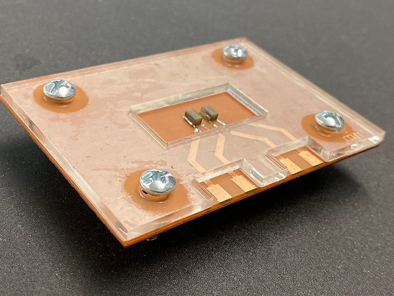
 A device uses sound waves to detect the stiffness of an extracellular matrix, a structural network that contains cells. Changes in the stiffness of this structure can indicate the spread of disease. Image Credit: Purdue University photo/Kayla Wiles.
Tracking the changes that occur in this structure—known as the extracellular matrix—would offer another way to scientists to analyze the development of cancer. However, it is difficult to detect changes that occur in the extracellular matrix, without damaging it.
Now, engineers at Purdue University have developed a new device that would enable disease specialists to place a sample of extracellular matrix onto a platform and identify its stiffness via sound waves.
The device has been demonstrated in a YouTube video and illustrated in a study published in the Lab on a Chip journal.
It’s the same concept as checking for damage in an airplane wing. There’s a sound wave propagating through the material and a receiver on the other side. The way that the wave propagates can indicate if there’s any damage or defect without affecting the material itself.
Rahim Rahimi, Assistant Professor, School of Materials Engineering, Purdue University
Rahimi’s laboratory creates novel biomedical devices and materials to deal with challenges relating to health care.
A unique extracellular matrix is associated with each organ and tissue, just like how street buildings differ in structure based on their purpose. Likewise, the extracellular matrix comes with “landlines,” or chemical and structural cues, that promote communication between separate cells accommodated in the matrix.
Although scientists have attempted to compress, stretch, or apply chemicals to extracellular matrix samples to quantify this environment, these techniques are also likely to affect the extracellular matrix.
A nondestructive way was eventually developed by Rahimi’s research team to analyze how the extracellular matrix reacts to therapeutic drugs, diseases, or harmful substances. For this research, the preliminary work was carried out in association with the laboratory of Sophie Lelièvre, a professor of cancer pharmacology at Purdue University, to find out how risk factors have an impact on the extracellular matrix and how they raise the risk of developing breast cancer.
The “lab-on-a-chip” device is linked to a receiver and transmitter. Once the extracellular matrix and the cells it contains are poured onto the platform, the transmitter produces an ultrasonic wave that travels via the material and subsequently activates the receiver. The result is an electrical signal that denotes the stiffness of the extracellular matrix.
The scientists initially showed the new device as a proof-of-concept with cancer cells held in a hydrogel. Hydrogel is a material that has a consistency analogous to an extracellular matrix. The scientists are currently analyzing the effectiveness of the device on collagen extracellular matrices.
Rahimi stated that the new device could be easily improved to run several samples simultaneously, such as in an array. This would enable scientists to look at many different aspects of a disease at the same time.
The study was carried out in Birck Nanotechnology Center in Purdue University’s Discovery Park and funded by the Breast Cancer Research Program, a Congressionally Directed Medical Research Program (grant W81XWH-17-1-0250).
Lab-on-a-chip Ultrasonic Platform to Monitor Tissue Culture Stiffness
The stiffening of a structure surrounding cells in the human body can indicate that cancer is invading other tissue. Being able to monitor changes to this structure, called the extracellular matrix, would give researchers another way to study the progression of disease. Video Credit: Purdue University/Erin Easterling.