Jun 3 2021
A research group from KTH has designed nano-sized particles, in an innovative way, to enhance the detection of tumors present inside the body and in biopsy tissue. The progress could allow early-stage tumors to be identified with lower doses of radiation.
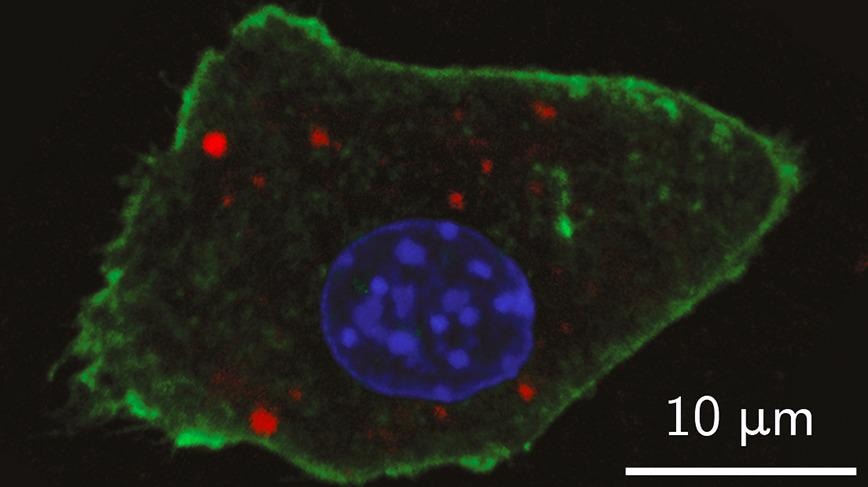
 A lab image shows the detection of the newly-developed nanoparticle contrast agents inside a mouse cell with optical fluorescence (in red). The cell nucleus and plasma membrane are depicted in blue and green, respectively. Image Credit: Giovanni Marco Saladino.
For visual contrast of living tissues to be improved, advanced imaging depends on agents like biomolecules and fluorescent dyes.
Progress in nanoparticle studies has extended the range of potential contrast agents for more targeted diagnostics, and currently, a research group from the KTH Royal Institute of Technology has increased the bar further. They have integrated X-ray and optical fluorescence contrast agents into an individual enhancer for both modes.
Muhammet Toprak, Professor of Materials Chemistry at KTH, stated that the production of contrast agents offers a new dimension in the field of X-ray bio-imaging. The study was published in ACS Nano, an American Chemical Society journal.
This unique design of nanoparticles paves the way for in vivo tumor diagnostics, using X-ray fluorescence computed tomography (XFCT).
Muhammet Toprak, Professor of Materials Chemistry, KTH Royal Institute of Technology
Toprak stated that the new 'core-shell nanoparticles' might play a role in the advent of theranostics, a portmanteau for diagnostics and therapy, in which, for instance, single drug-loaded particles have the potential to both detect and treat malignant tissues.
Shell Structure
The name core-shell contrast agent is derived from its architecture. It comprises a core combination of nanoparticles with earlier-established potential in X-ray fluorescence imaging, like molybdenum (IV) oxide and ruthenium.
This core is wrapped in a shell composed of silica and Cy5.5, a near-infrared fluorescence-emitting dye utilized for optical imaging methods like optical spectroscopy and microscopy.
Toprak states that the agent’s brightness is enhanced and its photo-stability is extended by encapsulating the Cy5.5 dye inside the silica shell, thereby allowing the X-Ray or dual optical imaging method. Besides, silica offers the advantage of tempering the toxic impacts of the core nanoparticles.
Improving the Odds of Detection
Tests performed with laboratory mice have demonstrated that the XFCT contrast agents allow the location of early-stage tumors measuring just a few millimeters in size.
According to Toprak, the technology paves the way to determine early-stage tumors present in the living tissue.
That is due to the existence of multiple contrast agents increasing the likelihood that diseased areas will appear in scans, even as the distribution of the nanoparticles turns out to be concealed by their communication with proteins or other biological molecules.
Nanoparticles of different size, originating from the same material, don’t appear to be distributed in the blood in the same concentrations. That’s because when they come into contact with your body, they’re quickly wrapped in various biological molecules—which gives them a new identity.
Muhammet Toprak, Professor of Materials Chemistry, KTH Royal Institute of Technology
Toprak states a multitude of contrast agents for XFCT would allow studying the biodistribution of nanoparticles in-vivo with the help of low-dose X-rays. That would enable determining the best size and surface chemistry of the nanoparticles for the preferred targeting and imaging of the diseased area.
Besides Toprak, the study co-authors included Giovanni M. Saladino, Carmen Vogt, Yuyang Li, Kian Shaker, Bertha Brodin, Martin Svenda, and Hans M. Hertz. The study was financially supported by the Knut and Alice Wallenberg’s Foundation, Grant number KAW2016.0057.
Journal Reference:
Saladino, G. M., et al. (2021) Optical and X-ray Fluorescent Nanoparticles for Dual Mode Bioimaging. ACS Nano. doi.org/10.1021/acsnano.0c10127.