With the growing demand for new applications in tissue regeneration, the functionalization of melt electrowritten scaffolds has gained the attention of researchers in recent years to fulfill the needs of tissue engineering. In one such work published in the journal of Nano Letters, the authors reported two types of surface modifications on melt electrowritten polycaprolactone (PCL) grids.

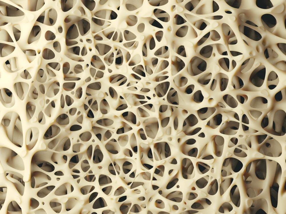
Study: Reshapable Osteogenic Biomaterials Combining Flexible Melt Electrowritten Organic Fibers with Inorganic Bioceramics. Image Credit: eranicle/Shutterstock.com
Here, they used zinc oxide (ZnO)-based nanomaterials to enhance osteogenic differentiation. The flexibility of PCL grids adds an advantage in obtaining reshapable osteogenic bioscaffold.
Surface Modification and Functionalization of Melt Electrowriting (MEW)
MEW enables the design and construction of high-resolution three-dimensional (3D) printing structures. Previous reports on MEW-based scaffolds like MEW PCL/hydroxyapatite (HAp), MEW PCL /hydrogel, mini lumbar vertebra-mimicking MEW discussed applications in bone tissue engineering. However, despite their extensive development, MEW scaffolds lack functionalization and are unsuitable for heat-labile molecules like proteins. Thus surface coating of MEW products is the most suitable way of surface modification and functionalization.
Due to the piezoelectric property of bones, they are highly regenerative and can heal faster on applying mechanical stress. In this context, incorporating ZnO-based nanoparticles was reported to trigger osteogenic differentiation and bone tissue engineering.
ZnO Nanomaterial Modified MEW PCL Grids
In the present study, for the first time, a group of researchers from Denmark developed two easy and reproducible strategies for ZnO-surface modifications on MEW PCL structures. The MEW PCL grid was surface modified with ZnO nanoparticles (NPs) and ZnO nanoflakes (NFs). They also surface-modified MEW PCL with HAp NPs for comparison.
The constructed modified MEW PCL grid scaffolds, in short names PCL/ZP with ZnO NPs and PCL/ZF with ZnO NFs in MC3T3 proliferation and differentiation, enabled further investigations. The results showed an increased alkaline phosphatase (ALP) expression and calcium deposition in PCL/ZP compared to PCL/HP and unmodified MEW PCL grid scaffolds, suggesting scaffold modification with ZnO NPs can enhance osteogenic properties.
Research Findings
The PCL and PCL/ZP grid scaffolds characterized by atomic force microscopy (AFM) showed that MEW PCL has a diameter of 10.0 ± 3.2 micrometers with a space gap of 200 micrometers.
Moreover, the scanning electron microscopy (SEM) images revealed the surface pattern of the pure PCL grid. It has polymer-based semicrystalline line pattens with nanoroughness.
The PCL, PCL/ZP 0.1 (PCL/ZP with 0.1 milligrams per milliliter ZnO NPs), and PCL/ZP 0.5 (PCL/ZP with 0.5 milligrams per milliliter ZnO NPs) grid scaffolds were biocompatible. In contrast, the PCL/ZP 1 (PCL/ZP with 1 milligram per milliliter ZnO NPs) grid scaffolds were cytotoxic. Furthermore, conducting a Cell Counting Kit-8 (CCK8) assay to evaluate the effect of ZnO NPs on MC3T3 cell viability showed that the presence of ZnO has a cytotoxic effect even in low doses in long-term cell culture.
The ALP activity of cells on PCL/ZP 0.1 and PCL/ZP 0.5 scaffolds was higher than pure PCL, indicating the enhanced activity due to ZnO coating.
The side view of PCL/ZF 6 (PCL grids after growth of ZnO NFs for six hours) grid scaffolds showed a thin layer of ZnO NFs, with a maximum NF length of approximately 2.37 micrometers. After 12 hours, the ZnO NFs grew only on one side of PCL grids to form a homogenous coating. The PCL/ZF grids mimic the flake-shaped hydroxyapatite nanocrystals surrounding collagen fibers of bone. It is important to note that the PCL/ZF 12 (PCL grids after growth of ZnO NFs for 12 hours) still showed flexibility for reshaping after ZnO NF coating.
Before cell seeding, the PCL/ZF scaffolds were pre-soaked in a growth medium for cell biocompatibility. Due to the high ZnO NF dose, PCL/ZF 12 showed more cytotoxicity than the tricalcium phosphate (TCP) group. The thick ZnO NFs coating on PCL grids enhanced the cell seeding due to the bigger diameter of PCL/ZF12 fibers.
The ALP activity levels were approximately 2.35 times higher in PCL/ZP 0.5 compared to PCL/HP 5 in week 5. Moreover, the authors reported achieving better results with ZnO NFs than with ZnO NPs. While the highest ALP activity was observed in samples of PCL/ZF 6 with approximately 2.11 times higher activity than PCL/ZP 1, the calcium mineralization in PCL/ZF 12 is approximately 2.17 times higher than PCL/ZP 0.1.
Conclusions and Future Scope
Through the present work, the authors have successfully modified MEW PCL scaffolds with piezoelectric ZnO to induce MC3T3 cell osteogenic differentiation. The reported surface microdissolution adhesion method in ethanol is a versatile approach for the facile application of other functional nanomaterials.
As ZnO NFs have restricted growth on the side of scaffolds, the side growth method serves best for MEW microporous scaffolds. ZnO NF enhanced osteogenic performance over ZnO NPs due to their biomimicking topography. Further, due to the MEW PCL grid’s flexibility, the PCL/ZF retained their reshapability.
The present work has great potential to increase the functionality of MEW scaffolds to widen their applications. Especially designing and synthesizing MEW PCL grid scaffolds modified with ZnO contributed to bone regeneration investigations.
Reference
Su, Y., Müller, C. A., Xiong, Xuya, Dong, M., and Chen, M. (2022). Reshapable osteogenic biomaterials combining flexible melt electrowritten organic fibers with inorganic bioceramics. Nano Letters. https://pubs.acs.org/doi/10.1021/acs.nanolett.1c04995
Disclaimer: The views expressed here are those of the author expressed in their private capacity and do not necessarily represent the views of AZoM.com Limited T/A AZoNetwork the owner and operator of this website. This disclaimer forms part of the Terms and conditions of use of this website.