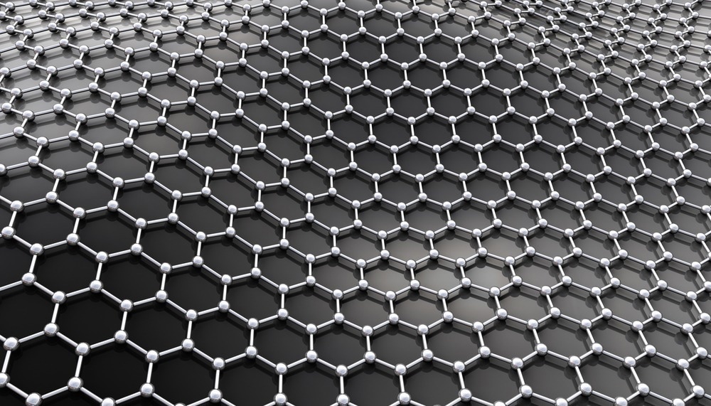
The purpose of this article is to provide readers with a deeper understanding of the state-of-the-art technology and development surrounding in situ graphene liquid cells-TEM.

Image Credit: Tatiana Shepeleva/Shutterstock.com
Liquid transmission electron microscopy (TEM) is one of the most powerful techniques for imaging a live reaction in a liquid state with reasonable resolution. Graphene's high impermeability, mechanical strength, and chemical inertness make it a perfect material for enclosing liquid solutions and imaging within situ transmission electron microscopy.
Early Strategies for Liquid TEM
To image liquid samples in TEM, the sample has to be sealed in a transparent cell. In early 2000 silicon nitride (SiN) was the material of choice for liquid encapsulation. However, this material usually results in a thicker liquid cell, and the range of the spatial resolution was only 10 nm.
In 2012, a graphene liquid cell was developed to overcome the resolution limit of the nitrite-based cell. This technique involves sealing a small amount of liquid (max. ~10-18 L) between two layers of single-layer graphene.
Under the influence of van der Waals forces, the top and bottom graphene flakes gradually come into contact, entrapping liquid within the well regions. Atomic resolution imaging (1 nm) was made possible by using graphene as an encapsulation rather than silicon nitride because of the thinness of the graphene membrane (0.34 nm).
Graphene as a Liquid Container
The benefits of graphene liquid cells are attributed to the unique properties of graphene. Graphene is one atom thick (0.34 nm), flexible, and electron transparent. As a result, incident electrons pass through graphene with minimal energy loss. Furthermore, graphene is extremely strong, both elastically and mechanically, and its membranes can withstand high pressures.
Second, because graphene is physically and theoretically impermeable to all atoms larger than a proton, it completely isolates your liquid inside the vacuum chamber.
Third, when an electron beam hits the liquid in a liquid cell, water can easily break down into reactive species. These reactive species can interfere with reactions in liquid cells or damage your sample. However, these radicals attach to the graphene layer in graphene liquid cells, reducing the radical effect.
Finally, because graphene is a good conductor of heat, image drifting from electron charging can be minimized.
Research Examples of Graphene Liquid Cell Application
Researchers from the University of California, Berkeley, were the first to report graphene liquid cells in 2012. The platinum salt precursors were encapsulated in graphene liquid cells in this study, allowing for real-time video and imaging of the early stages of nucleation and growth of platinum nanoparticles with very high spatial resolution.
Graphene liquid cells -TEM is also versatile, allowing researchers to study the real-time formation of bimetallic nanoparticles with complex morphologies such as hollow nanoparticles and core-shell structures.
The TEM technique with graphene liquid cells can be useful in understanding the battery reactions of various active materials, such as lithium-ion batteries. Making a circuit in graphene liquid cells is relatively difficult.
However, using electron beam illumination, we can induce chemical initiation, which is similar to the actual battery charging mechanism. Researchers reported that by enclosing Si nanoparticles in LiPF6 electrolytes with graphene liquid cells, they could obtain a real-time analysis of the volume expansion of silicon (Si) nanomaterials or the development of solid electrolyte interface layer during their electrochemical reactions with lithium ions. As a result, in situ TEM with graphene liquid cells served as a useful foundation for the robust design of high-capacity Si anodes in lithium-ion batteries.
According to the researchers, graphene liquid cells can also be used to image the structure and dynamics of web biological samples in a liquid environment with nanoscale resolution. As biomolecules can freely move inside graphene liquid cells, the movement and life reaction of the biomolecule can be observed.
Challenges and Ideas for Improvement with Graphene Liquid Cells
When placed in the TEM's vacuum, graphene liquid cells may experience some liquid sample evaporation due to gaps in the graphene flakes. To address this, it is suggested that defect-free high-quality CVD-grown graphene be used instead of exfoliated graphene.
As graphene sheets are generally hydrophobic, they are nonwetting for water. In this case, increasing the viscosity of the liquid may be an option.
The main experimental challenge is creating liquid pockets with a sufficient amount of liquid in a reproducible manner. Because the formation of these liquid pockets is random, their location and dimensions are unpredictable. To address this issue, scientists from the University of Manchester demonstrated a new graphene liquid cell design that consists of a thin lithographically patterned hexagonal boron nitride crystal encapsulated on both sides with graphene windows. This method provides unprecedented control over cell dimensions.
It has also been suggested that rough handling of graphene and the substrate can be avoided; instead of acid etching, bubbling and hot-water delamination methods for separating metal foil supporting graphene, be used for clean transfer of graphene to TEM grids.
References and Further Reading
Yuk, Jong Min, et al. (2012). High-resolution EM of colloidal nanocrystal growth using graphene liquid cells. Science. doi/10.1126/science.1217654
Yuk, Jong Min, et al. (2014). Anisotropic lithiation onset in silicon nanoparticle anode revealed by in situ graphene liquid cell electron microscopy. ACS nano. doi/10.1021/nn502779n
Wang, Canhui, et al. (2014). High‐resolution electron microscopy and spectroscopy of ferritin in biocompatible graphene liquid cells and graphene sandwiches. Advanced Materials. doi/full/10.1002/adma.201306069
Textor, Martin et al. (2018). Strategies for preparing graphene liquid cells for transmission electron microscopy. Nano letters. doi/10.1021/acs.nanolett.8b01366
Park, Jungjae, et al. (2021). Graphene liquid cell electron microscopy: Progress, applications, and perspectives. ACS nano. doi/10.1021/acsnano.0c10229
Kelly, Daniel J., et al. (2018). Nanometer resolution elemental mapping in graphene-based TEM liquid cells. Nano letters. doi/full/10.1021/acs.nanolett.7b04713
Disclaimer: The views expressed here are those of the author expressed in their private capacity and do not necessarily represent the views of AZoM.com Limited T/A AZoNetwork the owner and operator of this website. This disclaimer forms part of the Terms and conditions of use of this website.