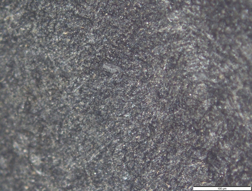
High temperature microscopy is the use of microscopy techniques on samples at high temperatures. Often, this means the microscopy instrumentation needs to be specifically designed to cope with the effects of high temperatures. This is partly because many optical components undergo temperature-dependent changes in their optical properties.

Image Credit: Maniock/Shutterstock.com
One of the key applications of high temperature microscopy is to examine material behavior under high temperature conditions. For example, high temperature microscopy can be used to understand the ferric grain formation sites in steel alloys and some of the material behavior associated with the phase transitions seen when the temperature is changed.
Nanoscience Applications
In the nanosciences, often high temperature microscopy methods make use of electron-based microscopies rather than optical experiments. This is because the nanoscale structures often demand a spatial resolution beyond what is achievable with most mainstream optical methods. As well for applications on metal alloys, high temperature microscopy can be used to study phenomena such as high temperature crystallization processes and the thermal stability of different material types, including glass-ceramics. Such studies can be performed over longer thermal aging timescales to ensure there is no degradation of the material properties or performance after extended periods of heating.
Often, phase changes in nanomaterials are of particular interest in high temperature microscopy stages. The nanoscale structural changes that occur during a phase transition are often of particular interest as there is often unusual physical behavior at grain boundaries and these nanoscale changes map into the overall macroscopic properties of the material.
Nanomaterials to be used for high temperature applications may undergo many phase changes during heating and cooling cycles. As a result, it is important that tiny, nanoscale or microscale defects do not form and lead to macroscopic material failure. High temperature microscopy can help identify such regions and the types of cracks that are forming with a view to manufacturing better materials. Inspection of small scale weld cracking to understand the best processing methods is another application of high temperature microscopy.
Microscopy Instrumentation
High-temperature microscopy is sometimes otherwise known hot-stage or temperature-controlled microscopy, where a heated stage is used to increase the sample temperature and a number of different measurements and sample analyses are performed.
Hot-stage microscopy can involve a full analysis of the sample of interest using methods such as differential scanning calorimetry (DSC) and thermomechanical analysis (TMA).
Hot-stage microscopy can be used in the analysis of pharmaceuticals, where using microscopy to observe the crystallization behavior of the active pharmaceutical ingredient under temperature ramping conditions can be used to determine the thermal stability of the active compound.
Access to methods such as DSC and TMA combined with optical imaging information can also be used to study small-scale polymorphism in detail, as different polymorphs of a pharmaceutical compound may differ in their physicochemical properties.
A typical hot-stage instrument will consist of the microscopy apparatus, a computer-controllable hot stage, any necessary filters and a camera to record the image information. Often, translation stages will be used to control the sample position or allow for rastering over the sample area. Some instrumentation also comes with cooling, as well as heating capabilities for recording a wider range of temperature conditions.
Most high temperature microscopes have a heating stage with a crucible capable of providing intense temperatures in the local sample environment. Depending on the heating process to be studied, the heating process may be done with an electrical heater or, in some cases, a laser can be used to initiate the heating.
The advantage of using a microscope alongside traditional thermal analysis methods like TMA or DSC is that as well as the latter being used to determine the phase transition points, the imaging information can be used to analyze any physical changes in the structure of the sample. Sometimes, when the thermal changes associated with a phase transition are very small, it is easier to identify the phase transition temperatures by visual inspection of the sample than through the results of methods like DSC.
Many microscopes make use of polarizers and filters to improve image contrast and quality. In electron microscopes, this tends to be done by using special electrostatic lenses to shape or change the focusing of the electron beam. Using brighter electron guns can help improve the levels of contrast in the images as well

Image Credit: S. Singha/Shutterstock.com
Recent Work
Nanoscale materials engineering, the pharmaceutical industry and, more recently, even the food industry are some of the key application areas for hot-stage microscopy. Recent work has involved the use of hot stage microscopes to look at how nanoparticle dispersion in liquid crystals changes as a function of temperature and what types of phase change the material undergoes during the heating and cooling cycles.
Thermoanalytical analysis of nanomaterials and pharmaceuticals is not just used for material analysis but also for understanding how drug delivery will work. Many therapeutics rely on atomic-level interactions between the therapeutic target and the therapeutic molecule.
High resolution microscopy can investigate how these small-scale changes are influenced by temperature and whether there are temperatures at which compounds start to undergo some kind of breakdown.
The food industry also exploits nanoscale effects when designing new food additives or preservatives, as these rely on the food-body interactions that are similar to therapeutics. Stabilization of particular food phases, such as foams, relies on these very small-scale interactions that new compounds look to exploit.
Overall, hot-stage microscopy is a highly versatile technique that offers a great deal of insight into material properties. While the spatial resolution is not as good as other standard microscopy methods, the ability to capture temperature-dependent behavior is very important for any material that will experience a range of temperature conditions in its application.
References and Further Reading
Mu, W., Hedstrom, P., Sibata, H., Jonsson, P. G., & Nakajima, K. (2018). High-Temperature Confocal Laser Scanning Microscopy Studies of Ferrite Formation in Inclusion-Engineered Steels : A Review. Advanced Real Time Optical Imaging, 70(10), pp. 2283–2295. https://doi.org/10.1007/s11837-018-2921-1
Gödeke, D., & Dahlmann, U. (2011). Study on the crystallization behaviour and thermal stability of glass-ceramics used as solid oxide fuel cell-sealing materials. Journal of Power Sources, 196, pp. 9046–9050. https://doi.org/10.1016/j.jpowsour.2010.12.054
Molinaro, C., Béné, M., Gorlas, A., Cunha, V. Da, Robert, H. M. L., Catchpole, R., Gallais, L., Forterre, P., & Baffou, G. (2022). Life at high temperature observed in vitro upon laser heating of gold nanoparticles. Nature Communications, 13, p. 5342. https://doi.org/10.1038/s41467-022-33074-6
Kumar, A., & Singh, P. (2020). Hot stage microscopy and its applications in pharmaceutical characterization. Applied Microscopy, 50, p. 12. https://doi.org/10.1186/s42649-020-00032-9
Patel, A. R. (2020). Functional and Engineered Colloids from Edible Materials for Emerging Applications in Designing the Food of the Future. Advanced Functional Materials, 30, p. 1806809. https://doi.org/10.1002/adfm.201806809
Cai, C., Cai, J., Zhao, L., & Wei, C. (2014). In situ Gelatinization of Starch using Hot Stage Microscopy. Food Sci Biotechnol, 23(1), pp. 15–22. https://doi.org/10.1007/s10068-014-0003-x
Disclaimer: The views expressed here are those of the author expressed in their private capacity and do not necessarily represent the views of AZoM.com Limited T/A AZoNetwork the owner and operator of this website. This disclaimer forms part of the Terms and conditions of use of this website.