Sep 10 2009
Researchers at The University of Nottingham have a new weapon in their arsenal of tools to push back the boundaries of science, engineering, veterinary medicine and archaeology.
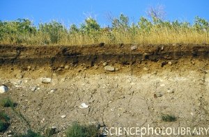
 Soil cross section. Credit: KENNETH W. FINK / SCIENCE PHOTO LIBRARY - © This image is for illustration only and subject to copyright and may not be used or copied in any way without prior permission from Science Photo Library http://www.sciencephotolibrary
From soils and sediments, to chunks of pavement, archaeological remains and chocolate bars... the Nanotom, the most advanced 3D X-ray micro Computed Tomography (CT) scanner in the world, will help scientists from a wide variety of departments across the University literally see through solids. The machine will make previously difficult and laborious research much easier as it allows researchers to probe inside objects without having to break into them.
The Nanotom has been supplied by GE Sensing and Inspection Technologies as part of a new project in the School of Biosciences to scan soil samples for research into soil- plant interactions. But it’s also an interdisciplinary piece of kit which will be used by other Schools for a wide variety of projects.
At least eight University departments will use the Nanotom. From scanning lichens in Biology, to sustainable building materials in the Built Environment, windblown sediments in Geography, and even animal muscle tissues in Veterinary Science, the machine will be a popular resource and at the moment, the only one of its kind at a British university. The Nanotom will also be hired out to private companies outside the University as a source of revenue that will help fund it.
CT is a very powerful tool that allows us to see the internal structure of an object that might be otherwise hidden from view. As well as its widespread use in healthcare, CT also has many applications in research and industry in the fields of Non-Destructive Testing (NDT) and Non-Destructive Evaluation (NDE) of solids. It’s used to carry out dimensional measurements, assembly checks, testing for the location and analysis of compositional defects. The University’s new machine will produce high-resolution 3D and slice images of solids with a pixel resolution of up to ½ micron or 500 nanometres.
The Nanotom will be based at the School of Biosciences as the centrepiece of research into efforts to understand the microscopic interactions between plant root growth and soil structure. The first project to use it will examine the sensing ability of roots to grow in the best direction for the health of the plant through the soil. It aims to provide evidence of how the root reacts and adapts to soil stresses like drought and compaction by adjusting the genetic information in the tips of the root as it grows. The Nanotom will allow researchers to follow the progress of the root growth and soil structural development for the first time without disturbing the sample of the plant growing in the soil.
The eventual aim of research like this is to contribute to worldwide efforts for food security and sustainable food production by preserving and improving the vital but finite soil resources of the planet. It will enable scientists to come up with a recipe for the best soil composition and level of compaction as well as informing plant breeding programmes. Accurate soil structure measurement will be also be essential in changing farming practices to cut CO2 which is released into the atmosphere during traditional ploughing of agricultural soil.
Dr Sacha Mooney from the University’s Division of Agricultural & Environmental Sciences, said: “This new kit will completely revolutionise our work in trying to understand the key factors that control some of the many functions that soils perform. The days of considering the soil, or any porous media, simply as a ‘black box’ are behind us. Now we can see into the fragile structures as they exist in the field and hopefully use the microscopic measurement of the complex 3-D structure to enhance and develop models concerning how soils behave. Of course it’s not just soils we’ll be scanning, I think I am just as excited about the opportunity to look inside newly created environmental building materials, eco-friendly crops developed to improve yield and even chocolate bars for the food industry!”
Pro-Vice-Chancellor for Research, Professor Bob Webb, said: “As a leading research university internationally, we are proud to be investing in this cutting-edge technology. The new scanner will be a vital resource across many departments and commercial research partners outside the University. It will enable faster, more accurate research into many scientific areas and we have no doubt it will bring real results in crucial projects like global food production.”