Oct 5 2009
A fiber-optic sensor created by a team of Purdue University researchers that is capable of measuring oxygen intake rates could have broad applications ranging from plant root development to assessing the effectiveness of chemotherapy drugs.
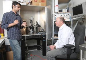
 Mohammad Rameez Chatni, at left, and Marshall Porterfield developed the self-referencing optrode that can measure oxygen intake in real time. (Credit: Purdue Agricultural Communications photo/Tom Campbell)
The self-referencing optrode, developed in the lab of Marshall Porterfield, an associate professor of agricultural and biological engineering, is non-invasive, can deliver real-time data, holds a calibration for the sensor's lifetime and doesn't consume oxygen like traditional sensors that can compete with the sample being measured. A paper on the device was released on the early online version of the journal The Analyst this week.
"It's very sensitive in terms of the biological specimens we can monitor," Porterfield said. "We don't only measure oxygen concentration, we measure the flux. That's what's important for biologists."
Mohammad Rameez Chatni, a doctoral student in Porterfield's lab, said the sensor could be used broadly across disciplines. Testing included tumor cells, fish eggs, spinal cord material and plant roots.
Cancerous cells typically intake oxygen at higher rates than healthy cells, Chatni said. Measuring how a chemotherapy drug affects oxygen intake in both kinds of cells would tell a researcher whether the treatment was effective in killing tumors while leaving healthy cells unaffected.
Plant biologists might be interested in the sensor to measure oxygen intake of a genetically engineered plant's roots to determine its ability to survive in different types of soil.
"This tool could have applications in biomedical science, agriculture, material science. It's going across all disciplines," Chatni said.
The sensor is created by heating an optical fiber and pulling it apart to create two pointed optrodes about 15 microns in diameter, about one-tenth the size of a human hair. A membrane containing a fluorescent dye is placed on the tip of an optrode.
Oxygen binds to the fluorescent dye. When a blue light is passed through the optrode, the dye emits red light back. The complex analysis of that red light reveals the concentration of oxygen present at the tip of the optrode.
The optrode is oscillated between two points, one just above the surface of the sample and another a short distance away. Based on the difference in the oxygen concentrations, called flux, the amount of oxygen being taken in by the sample is calculated.
It's the intake, or oxygen transportation, that Porterfield said is important to understand.
"Just knowing the oxygen concentration in or around a sample will not necessarily correlate to the underlying biological processes going on," he said.
Porterfield said future work will focus on altering the device to measure things such as sodium and potassium intake as well. The National Science Foundation funded the research.