Sep 28 2010
Getting an inside look at the center of a cell can be as easy as a needle prick, thanks to University of Illinois researchers who have developed a tiny needle to deliver a shot right to a cell's nucleus.
Understanding the processes inside the nucleus of a cell, which houses DNA and is the site for transcribing genes, could lead to greater comprehension of genetics and the factors that regulate expression. Scientists have used proteins or dyes to track activity in the nucleus, but those can be large and tend to be sensitive to light, making them hard to use with simple microscopy techniques.
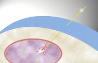
 University of Illinois researchers developed a nanoneedle that releases quantum dots directly into the nucleus of a living cell when a small electrical charge is applied.
Researchers have been exploring a class of nanoparticles called quantum dots, tiny specks of semiconductor material only a few molecules big that can be used to monitor microscopic processes and cellular conditions. Quantum dots offer the advantages of small size, bright fluorescence for easy tracking, and excellent stability in light.
"Lots of people rely on quantum dots to monitor biological processes and gain information about the cellular environment. But getting quantum dots into a cell for advanced applications is a problem," said professor Min-Feng Yu, a professor of mechanical science and engineering.
Getting any type of molecule into the nucleus is even trickier, because it's surrounded by an additional membrane that prevents most molecules in the cell from entering.
Yu worked with fellow mechanical science and engineering professor Ning Wang and postdoctoral researcher Kyungsuk Yum to develop a nanoneedle that also served as an electrode that could deliver quantum dots directly into the nucleus of a cell – specifically to a pinpointed location within the nucleus. The researchers can then learn a lot about the physical conditions inside the nucleus by monitoring the quantum dots with a standard fluorescent microscope.
"This technique allows us to physically access the internal environment inside a cell," Yu said. "It's almost like a surgical tool that allows us to 'operate' inside the cell."
The group coated a single nanotube, only 50 nanometers wide, with a very thin layer of gold, creating a nanoscale electrode probe. They then loaded the needle with quantum dots. A small electrical charge releases the quantum dots from the needle. This provides a level of control not achievable by other molecular delivery methods, which involve gradual diffusion throughout the cell and into the nucleus.
"Now we can use electrical potential to control the release of the molecules attached on the probe," Yu said. "We can insert the nanoneedle in a specific location and wait for a specific point in a biologic process, and then release the quantum dots. Previous techniques cannot do that."
Because the needle is so small, it can pierce a cell with minimal disruption, while other injection techniques can be very damaging to a cell. Researchers also can use this technique to accurately deliver the quantum dots to a very specific target to study activity in certain regions of the nucleus, or potentially other cellular organelles.
"Location is very important in cellular functions," Wang said. "Using the nanoneedle approach you can get to a very specific location within the nucleus. That's a key advantage of this method."
The new technique opens up new avenues for study. The team hopes to continue to refine the nanoneedle, both as an electrode and as a molecular delivery system.
They hope to explore using the needle to deliver other types of molecules as well – DNA fragments, proteins, enzymes and others – that could be used to study a myriad of cellular processes.
"It's an all-in-one tool," Wang said. "There are three main types of processes in the cell: chemical, electrical, and mechanical. This has all three: It's a mechanical probe, an electrode, and a chemical delivery system."
The team's findings will appear in the Oct. 4 edition of the journal Small. The National Institutes of Health and the National Science Foundation supported this work.
Source: http://illinois.edu/