Whenever a person experiences a heart attack, a portion of the heart stops working and its nerve cells and special cells that contract and expand on their own accord are lost forever. This damaged area could not be repaired by surgeons and instead they abandon it.
A group of researchers from India’s Indian Institute of Technology Kanpur and from the Brown University focused on this problem and tried to resuscitate the deadened area. They resorted to nanotechnology and assembled a structure resembling a scaffold called nanopatch, made up of carbon nanofibers along with a polymer, which is approved by the government. Further testing revealed that the synthetic nanopatch restored and revived the natural heart tissue cells named cardiomyocytes. David Stout, a graduate student at Brown University’s School of Engineering and who is also the chief author of a research paper which has been published in Acta Biomaterialia, has revealed that the main idea was to place this patch on the area where there was dead tissue so that it could be regenerated and help to develop a healthy heart.
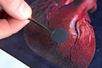
 Engineers at Brown University have created a nanopatch for the heart that tests show restores areas that have been damaged, such as from a heart attack.
According to the American Heart Association in the year 2009, over 785,000 Americans suffered from a new kind of heart attack connected to weakness resulting from scarred cardiac muscle from a prior heart attack. The Association also reveals that a fifth of the men and a third of the women who have gone through a heart attack would probably suffer from another one inside six years. If the new approach proves successful, then it would be a boon for millions of people.
The researchers at both the Indian Institute of Technology, Kanpur and at the Brown University utilized carbon nanofibers which were helical shaped tubes with a diameter ranging between 60 and 200 nm. The engineers found that the nanofibers being excellent conductors of electrons performed excellently the kind of electrical connections which the heart needs for beating at a steady rate. They created a 22 mm long mesh by sewing the nanofibers together with the help of a poly lactic-co-glycolic acid polymer emulating a black band aid. They placed the mesh on a glass substrate to see if cardiomyocytes would colonize or inhabit the surface and help the growth of more cells. The tests revealed that after four hours the nanofibers seeded with cardiomyocytes five times more than the control sample which consisted of just the polymer alone. After a period of five days the density was reportedly six times more than that of the control sample and also had double the neuron density after four days.
According to Thomas Webster, who is the Associate Professor in Orthopedics and Engineering at the Brown University and also one of the authors of the research paper, the scaffold being highly durable and elastic could easily contract and expand just like the heart tissue thus helping the neurons and the cardiomyocytes to assemble on the scaffold and produce new cells and regenerate the area. The researchers would further like to nip the scaffold pattern to ape the electrical current of the heart and construct an in-vitro mock-up, with which tests could be carried out to see how the material responds to the heart’s beat and voltage system. They also wanted to make certain that the cardiomyocytes that develop on the scaffolds have the same abilities as other heart tissue cells.