Mar 2 2013
A professor in the Department of Electrical and Computer Engineering at Texas A&M University and his Ph.D. student recently received a best paper award from a prestigious conference on optics and photonics.
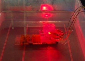
 MEMS mirror in scanning motion under water
Associate Professor Jun Zou and Chih-Hsien Huang were among the co-authors to receive the Seno Medical Best Paper Award at the 2013 SPIE Photonic West Conference in San Francisco. Their paper, “Water-immersible MEMS scanning mirror designed for wide-field fast-scanning photoacoustic microscopy,” is a joint work between Zou’s Micro/Nano Imaging and Sensing Technologies (MIST) group and Dr. Lihong Wang’s Optical Imaging Lab in the biomedical engineering department at Washington University in St. Louis.
Photo of MEMS mirror in scanning motion under waterMEMS (micro-electromechanical systems) scanning mirrors have been extensively developed for optical imaging, communication and projection. A typical example is the Texas Instruments’ digital mirror device (DMD) used in the DLP overhead projectors, projection TVs and digital cinemas. However, just like a mosquito getting trapped in water or oil, existing MEMS scanning mirrors will fail when immersed in liquids. By creating new designs and fabrication processes, Zou’s group has overcome this limitation. With the water-immersible MEMS scanning mirror, it is now possible to achieve fast and simultaneous scanning of not only optical beams but also high-frequency ultrasound waves (which can only travel in liquids or solids) in a liquid environment. (Pictured left: MEMS mirror in scanning motion under water)
Photoacoustic microscopy (PAM) is a new imaging modality recently developed in Wang’s lab, which utilizes focused optical and ultrasound waves to interrogate live cells and tissues even without additional contrast agents. However, limited by the slow speed of the conventional scanning components, existing PAM systems usually take minutes to acquire a snapshot. With the new water-immersible MEMS scanning mirror, the imaging time is reduced to seconds or a fraction of a second. This turns the PAM system from a still camera into a high-speed camcorder. Now it is possible to capture Photo of tissue microvasculature imaged by PAMmany dynamic physiological processes and pathological conditions not observable before, which are otherwise important for the fundamental understanding of the functioning of human body and also the treatment of many acute human diseases, such as cancers, seizures and bacterial infections. In the future, the two teams will work together to further develop the new MEMS-enable PAM technology for both fundamental biomedical research and clinical diagnosis applications. (Pictured right: tissue microvasculature imaged by PAM)
Zou joined the Texas A&M electrical and computer engineering department in 2004 after the completion of his Ph.D. and postdoctoral training in electrical engineering at the University of Illinois at Urbana-Champaign. He is currently an associate professor in the solid-state device and nanoengineering area, where he leads the MIST group to advance novel imaging and sensing methods and systems to overcome ambiguous situations in the information era.
SPIE is the international Society for optics and photonics. SPIE Photonics Westis the most influential conference for biophotonics and biomedical optics, high-power laser manufacturing, optoelectronics, microfabrication and green photonics.