Dec 8 2017
DESY researchers have designed innovative lenses with the potential to perform X-ray microscopy with nanometer-scale resolution. The team headed by Saša Bajt, DESY scientist from the Center for Free-Electron Laser Science (CFEL), used innovative materials to perfect the design of exclusive X-ray optics and accomplished a focus spot size that had a diameter of less than 10 nm, where 1 nm is one-millionths of 1 mm and is smaller than the size of majority of the viruses. The study has been published in the Light: Science and Applications journal. The team has successfully applied the developed lenses to image marine plankton samples.
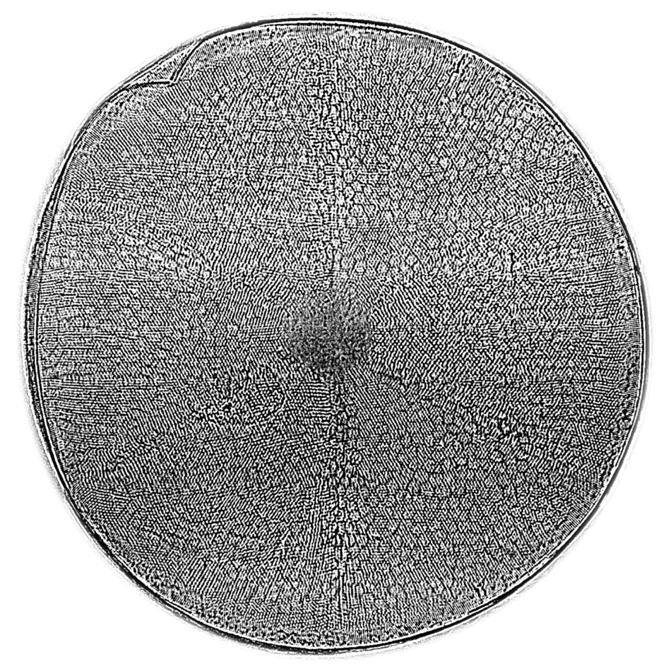
 The silica shell of the diatom Actinoptychus senarius, measuring only 0.1 mm across, is revealed in fine detail in this X-ray hologram recorded at 5000-fold magnification with the new lenses. The lenses focused an X-ray beam to a spot of approximately eight nanometers diameter—smaller than a single virus—which then expanded to illuminate the diatom and form the hologram. CREDIT: DESY/AWI, Andrew Morgan/Saša Bajt/Henry Chapman/Christian Hamm.
Present-day particle accelerators offer high-quality and ultra-bright X-ray beams. The penetrating nature and shorter wavelength of X-rays render them optimal for the microscopic analysis of complex materials. Yet, using these characteristics to their full benefits mandates extremely efficient and nearly perfect optics in the X-ray regime. Although globally there have been large-scale attempts, this has been very difficult than anticipated, and developing an X-ray microscope with the potential to enable a resolution of less than 10 nm is a very difficult task.
The distinctive characteristics of X-rays renders them difficult to be focus easily when compared to visible light. One technique to overcome this difficulty would be to use specialised X-ray optics known as multilayer Laue lenses, or MLLs, which include interspersed layers of two distinct materials with a thickness of few nanometers. They are developed by a coating technique known as sputter deposition. Unlike traditional optics, rather than refracting light, MLLs function by diffracting the incident X-rays in a manner that the beam is focused on a small spot. In order to accomplish this, the materials’ layer thickness must be accurately regulated. The thickness as well as orientation of the layers should be gradually altered throughout the lens. The focus size corresponds to the smallest layer thickness of the MLL structure.
In order to achieve the desired accuracy, Bajt and her colleagues integrated an innovative fabrication technique with in-depth knowledge of the material characteristics, which normally differ with respect to the layer thickness. The innovative lenses comprise more than 10,000 interspersed layers of an innovative combination of the materials silicon carbide and tungsten carbide. “The selection of the right material pair was critical for the success,” reiterates Bajt. “It does not exclude other material combinations but it is definitely the best we know now.”
If an X-ray beam has to be focused in the horizontal as well as vertical directions, it has to be passed through two perpendicularly placed lenses. Applying this framework, a spot size of 8.4 nm ´ 6.8 nm was achieved at the Hard X-ray Nanoprobe experimental station at the National Synchrotron Light Source NSLS II at Brookhaven National Laboratory, United States. The X-ray microscope’s resolution is governed by the focus size. The resolution of the innovative lenses is nearly five times more than that can be accomplished by using conventional ultra-modern lenses.
“We produced the world’s smallest X-ray focus using high efficiency lenses,” stated Bajt. Because of their penetrating character, X-rays will normally pass straight through the materials of lenses. It is quite apparent that such rays do not play a part in the focus, and hence a long-time aim has been to develop lens structures that improve the interaction with X-rays, to make a higher proportion of rays to be directed into the focus. The efficiency of the innovative lenses is over 80%. Such a high efficiency is accomplished by using the layered structures used to form the lens and which function similar to an artificial crystal and diffract X-rays in a regulated manner.
The higher efficiency accomplished in this case indicates the higher control level in developing the required nanometer structures. This precision enables projection imaging over a wide range of magnifications as showed by investigations of the innovative lenses. At beamline P11 of DESY’s X-ray source PETRA III, the team synthesized high-resolution holograms of Acantharea, single-celled Radiolaria belonging to the class of marine plankton and the only organisms with skeletons formed of the mineral strontium sulfate (SrSO4) or celestite.
Bajt’s and her colleagues have also applied the innovative lenses to take images of the biomineralized shells of marine planktonic diatoms. The shells of these single-celled organisms are very intricate, which are highly complex stable but also lightweight constructions. They comprise nanostructured silica, seen in two-dimensional investigations by using electron microscopes. Largely due to this structuring, silica’s strength is extremely high, of the order of 10 times greater when compared to construction steel, despite the fact that it is synthesized under low pressure and temperature conditions.
We hope that the novel X-ray optics will soon make it possible to image these nanostructures in 3D. This will enable us to model and understand the high mechanical performance of these shells and help us to develop new, environmentally friendly and high performance materials,.
Christian Hamm, Alfred Wegener Institute, Helmholtz Centre for Polar and Marine Research (AWI), who offered the samples and is one of the co-authors of this research.
The innovative lenses developed by the team can be applied used in a broad array of applications such as nano-resolution imaging and spectroscopy.
These MLLs open up new and exciting opportunities in X-ray science. They can be designed for different energies and used with coherent sources, such as X-ray free-electron lasers, this great achievement would not have been possible without a wonderful team with expertise in X-ray optics and theory, nanofabrication, material science, data processing and instrumentation. Since we now know how to optimise the lens design, our work paves the way to ultimately reach the goal of one nanometre resolution in X-ray microscopy.
Saša Bajt, DESY scientist from the Center for Free-Electron Laser Science (CFEL)
This study included researchers from DESY, the University of Hamburg, the National Science Foundation BioXFEL Science and Technology Center in the United States, Arizona State University in the United States, the University of Bialystok in Poland, Brookhaven National Laboratory in the United States, and Alfred Wegener Institute in Germany. CFEL is a cooperation of DESY, the University of Hamburg, and the German Max Planck Society.