Feb 28 2019
Using a new type of microscope, doctors may now be able to get a better idea of how safely and efficiently a pill will perform in the body.
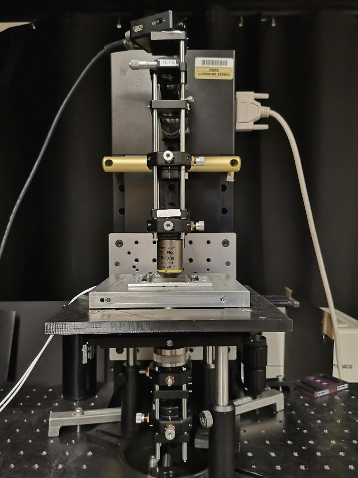
 A new type of microscope from Purdue University stacks the reference object and the one being examined on top of each other, instead of the conventional approach of having them side by side. (Image credit: Garth Simpson, Purdue University)
Researchers at Purdue University built the microscope founded on concepts of phase-contrast microscopy, which requires using optical devices to see membranes, molecules, or other nanoscale items that may be too translucent to distribute the light involved with conventional microscopes.
One of the problems with using the available microscopes or optical devices is that they require a point of reference for the scattered light, since the object being viewed is too optically transparent to scatter the light itself. We created a unique kind of microscope that stacks the reference object and the one being examined on top of each other with our device, instead of the conventional approach of having them side by side.
Garth Simpson, Study Lead and Professor of Analytical and Physical Chemistry, College of Science, Purdue University.
The Purdue microscope uses technology to interfere light from a featureless reference plane and a sample plane, quantitatively recovering the elusive phase shifts prompted by the sample. The work has been reported in the February 18th edition of Optics Express.
By just incorporating two small optics to the base design of a conventional microscope, the researchers were able to create their device. The change allows the team to collect better information and data about the object being studied.
The microscope we have created would allow for better testing of drugs. You could use our optical device to study how quickly and safely some of the active ingredients in a particular medication dissolve. They may crystallize so slowly that they pass through the body before dissolving, which significantly lowers their effectiveness.
Garth Simpson, Study Lead and Professor of Analytical and Physical Chemistry, College of Science, Purdue University.
Simpson said the microscope built at Purdue could also be employed for other forms of biological imaging, such as the ability to examine membranes and individual cells from the body for different medical testing.
Their effort aligns with Purdue’s Giant Leaps celebration, recognizing the university’s worldwide advancements in health as part of Purdue’s 150th anniversary. Health research, including progressive biological imaging, is one of the four topics of the yearlong celebration’s Ideas Festival, designed to promote Purdue as an intellectual center solving real-world problems.
Simpson has partnered closely with the Purdue Office of Technology Commercialization on several patented technologies including this one produced in his lab.