Dec 16 2019
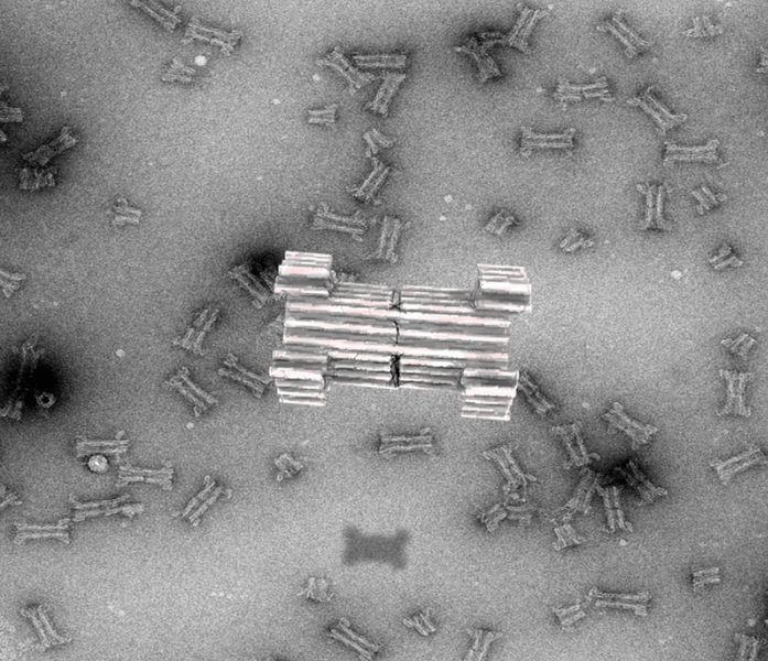
Headed by scientists from the iNANO/Department of Molecular Biology and Genetics at Aarhus University and the Department of Chemistry at the University of Copenhagen, a scientific collaboration has led to the development of a synthetic DNA nanopore that can selectively translocate protein-sized macromolecules across lipid bilayers.
 In the future, the newly discovered mechanism will potentially enable insertion of the sensor specifically into diseased cells and may allow diagnosis at the single cell level. Image Credit: Rasmus Peter Thomsen/AU.
Oxford Nanopore Technologies launched the first-ever commercial nanopore DNA sequencing device in 2015. Nanopore sequencing is based on a synthetically engineered transmembrane protein and enables channeling of long DNA strands through the pore’s central lumen, where changes in the ionic current act as a sensor of the individual bases in the DNA.
This method was a major landmark for DNA sequencing, and the success could be achieved only after several decades of research.
From that time, scientists have made efforts to extend this principle and develop larger pores to hold proteins that can be used for sensing purposes. However, these efforts have faced a major difficulty, which is the limited insight into artificial protein design.
As a substitute, a new method first reported by the AU group in 2009 has emerged. This method is based on artificial folding of DNA into complex structures, what is known as the 3D-origami method.
When compared to proteins, DNA origami has been demonstrated to have an unparalleled design space for developing nanostructures that mimic and extend naturally existing complexes.
In a new study, reported in Nature Communications, the scientists now describe the development of a large synthetic nanopore developed from DNA. This nanopore structure has the capability to translocate large protein-sized macromolecules between compartments isolated by a lipid bilayer.
Furthermore, a functional gating system was inserted into the pore to allow biosensing of very few molecules in solution.
The researchers used powerful optical microscopes, which enabled them to track the flow of molecules through each nanopore. The insertion of a controllable plug into the pore further enabled size-selective control of the flow of protein-sized molecules and demonstration of real-time, label-free biosensing of a trigger molecule.
Finally, a set of controllable flaps was attached to the pore. These flaps enabled targeted insertion into membranes that displayed specific signal molecules. In the years to come, this mechanism will possibly allow the insertion of the sensor particularly into diseased cells, and might enable diagnosis at the single cell level.