Feb 18 2021
Ever since X-rays were discovered by Wilhelm Röntgen in 1895, they have become a standard in medical imaging.
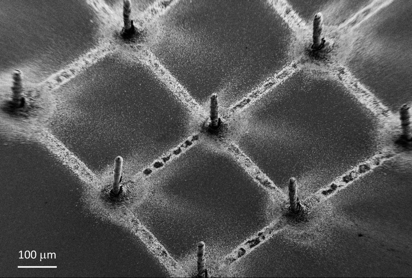
 Example of the deposited perovskite pillars, defining a pixel for the creation of an image. Image Credit: L. Forró, EPFL.
Less than one month after Röntgen’s well-known article was published, Connecticut–based doctors took the world’s first radiograph of a boy's broken wrist.
Since that time, there has been plenty of developments. Apart from radiographs, which the majority of individuals would have had at least once in their lifetime, today’s medical applications of X-rays include computer tomography (CT), fluoroscopy, and radiotherapy for cancer, in which numerous X-ray scans of the body are taken from various angles and these are subsequently integrated into a computer to produce virtual cross-sectional “slices” of the human body.
Nevertheless, medical imaging usually functions with low-exposure conditions, and hence needs high-resolution and cost-effective detectors that can work at the so-called “low photon flux.” Photon flux only explains the number of photons that strike the detector at a specified time and establishes the number of electrons that are consequently produced by the detector.
A research team, headed by László Forró from the School of Basic Sciences, has now designed this device unit. With the help of 3D aerosol jet-printing, the researchers created a new technique for generating extremely efficient X-ray detectors that can be effortlessly incorporated into typical microelectronics to dramatically enhance the performance of medical imaging devices.
These novel detectors are composed of graphene as well as perovskites—materials composed of organic compounds attached to a metal. The detectors are multipurpose, can be easily produced, and are at the vanguard of a broad range of applications, such as photodetectors, lasers, LED lights, and solar cells.
While aerosol jet-printing is quite new, it is nevertheless used for making 3D-printed electronic components, such as thin-film transistors, sensors, antennas, resistors, and capacitors, or for printing electronics on a specific substrate, for example, a cellular phone.
At the CSEM based in Neuchatel, the investigators used the aerosol jet printing device and then 3D-printed perovskite layers on a graphene substrate. The concept is that the perovskite within a device functions as both electron discharger and photon detector, while the outgoing electrical signal is amplified by graphene.
The researchers utilized the methylammonium lead iodide perovskite (MAPbI3), which has lately attracted a great deal of interest due to its intriguing optoelectronic characteristics, which pair well with its minimal production cost.
This perovskite has heavy atoms, which provide a high scattering cross-section for photons, and makes this material a perfect candidate for X-ray detection.
Endre Horváth, Chemist, School of Basic Sciences, EPFL
The outcomes were remarkable. The new technique created X-ray detectors that not only had a record sensitivity but also had four times enhancement on the most superior medical imaging devices.
By using photovoltaic perovskites with graphene, the response to X-rays has increased tremendously. This means that if we would use these modules in X-ray imaging, the required X-ray dose for forming an image could be decreased by more than a thousand times, decreasing the health hazard of this high-energy ionizing radiation to humans.
László Forró, School of Basic Sciences, EPFL
Another benefit of the perovskite-graphene detector is that it can be easily used to create images.
“It doesn’t need sophisticated photomultipliers or complex electronics. This could be a real advantage for developing countries,” Forró concluded.
Journal Reference:
Glushkova, A., et al. (2021). Ultrasensitive 3D Aerosol-Jet-Printed Perovskite X-ray Photodetector. ACS Nano. doi.org/10.1021/acsnano.0c07993.